Thomas H. Herdt DVM
Liver biopsy is a very useful way of assessing copper status in cattle, as well as for other diagnostic uses such as assessing liver fat concentration. In the past, rather large liver specimens were required for copper determination. The analytical equipment available today allows us to accurately measure copper and other mineral concentrations on very small liver samples. These samples can be taken quickly, conveniently, and with little or no risk to the animal.
The Tru-Cut type biopsy instrument is an economical and convenient means of collecting liver samples for copper analysis. There are several versions of the Tru-Cut instrument available. All are based on the same principle for cutting the sample, which is described further below. Entering “Tru-Cut” into the Google search engine will yield a variety of suppliers and variations on the basic instrument design. The needle pictured below is a simple and relatively inexpensive version of the Tru-Cut.

This needle may be purchased from Care Express Products (www.careexpress.com) in Cary, IL. The needles come in cartons of 10, and the current price may be checked at the Care Express website. The needles are designed for single use in human patients, but they may be cleaned and used repeatedly in veterinary applications. For bovine liver biopsy, the needle should be 14G and either four or six inches long. The four-inch needle will be of sufficient length in all but very well-finished beef animals.
These needles work by using the sharp beveled tip of the needle to cut a slice of liver into a notch in the stylet. The instrument is easy to use, but it is important that the actions of the needle and stylet be taken in the proper order, as described below.

Step 1. Insert the instrument into the animal with the stylet withdrawn into the needle, as shown. The site, direction, and depth of insertion are described further on.

Step 2. Advance the stylet into liver tissue as needle is held stationary. Liver tissue fills notch.

Step 3. Advance the needle while holding the stylet stationary. It is critical not to pull the stylet back because there is no cutting surface on the stylet. The sharp edge of needle point cuts the biopsy sample into notch.

Step 4. Advance the needle all the way over the stylet. This captures the sample and the instrument may be withdrawn from the animal.
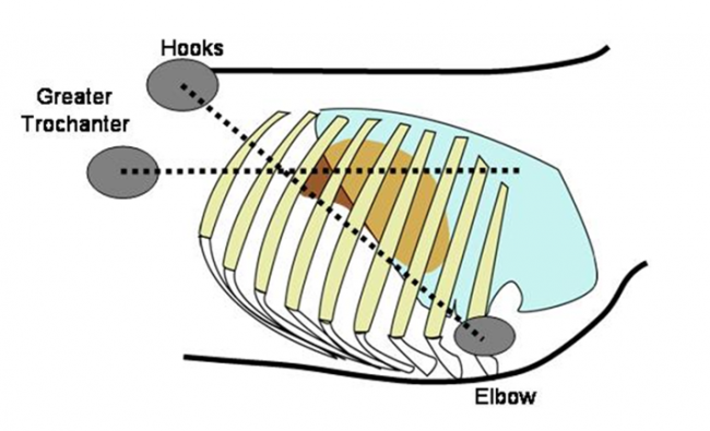
To identify the site at which to insert the needle, first identify the 10th intercostal space. Remember there are 13 ribs and 12 intercostal spaces, so the 10th intercostal space is the third from the last intercostal space. Then draw an imaginary line from the tuber coxae (hook) to the elbow. The point at which this line intersects the 10th intercostal space is the point at which to insert the needle. An alternative way to identify the site is to draw an imaginary line through the greater trochanter (hip) and parallel to the ground. The site at which this line intersects the 10th intercostal space may also be used as the landmark for needle insertion. Usually, there will be a space of about two inches within the 10th intercostal space and between these two imaginary lines. Anywhere within that space should be suitable for the biopsy site. The site of needle insertion is illustrated in the figure below.

The figure illustrates the site for insertion of the liver biopsy instrument. Note that the largest portion of the liver lies beneath the lung, leaving only a small portion of the caudal liver accessible directly through the body wall. The reflection of the diaphragm is actually caudal to the edge of the lung, making it necessary to penetrate the diaphragm to biopsy the liver via the 10 intercostal space.
The site should be prepared as if for surgery. It is useful to infiltrate 2 to 5 mL of 2% lidocaine solution under the skin and into the intercostal muscles at the site of insertion of the biopsy instrument. It is also useful to make a small stab incision through the skin in preparation for inserting the biopsy needle. A #15 Bard Parker blade is of convenient size for making a stab incision just big enough to accommodate the biopsy needle.
Insert the needle approximately parallel to the ground and angled slightly forward toward the left shoulder of the animal. The needle needs only to penetrate the skin, intercostal muscles, and diaphragm before entering the liver, a distance of only about two and one half inches in most adult cows. You will usually notice a distinct “popping” sensation as the needle punctures the diaphragm. You will also notice that the needle moves with the animal’s respirations after it is through the diaphragm. When the needle is in this position proceed with taking the biopsy, as described in the figures above.
With the needle held in place over the stylet (as in step 4 above), withdraw the instrument. Advance the stylet and observe how much of the notch is filled with liver tissue. If the notch is completely full, that is usually about 10 to 12 mg of liver tissue. For optimal copper analysis, we need at least 20 mg of liver tissue, meaning that it is usually necessary to take two samples.
Use the tip of a hypodermic needle to remove the liver sample from the instrument and transfer it to a test tube. It is best to use a “royal blue” stopper Vacutainer tube because these tubes are manufactured to be free of trace element contamination. These tubes are available from the MSU VDL, or from other suppliers of Vacutainer brand tubes. Using these tubes is particularly important if zinc is of concern because the rubber stoppers in most test tubes contain zinc and can thus lead to contamination of the sample.
The liver biopsy procedure is very safe, and no complications have been observed in hundreds of samplings. However, there is some small potential of inducing necrotic hepatitis, so if the animals have not been vaccinated against clostridial diseases, it is probably wise to administer an injection of penicillin or similar antibiotic at the time of sampling.
The test tubes containing the samples should be sent to the laboratory by overnight courier. Do not add formalin, saline, or anything else to the samples. It is not necessary to ship them on ice.
For herd evaluation, at least seven animals per feeding group should be tested. For optimal herd evaluation these should not be animals with obvious signs of infectious or inflammatory disease.
For current pricing for Minerals, Tissue or any other tests we offer, please see our catalog of available tests.
