By: Steve Bolin, DVM, MS, PhD
Vector-borne Diseases: Big Illnesses, Small Delivery Vehicles
From spring through fall in most of the United States, awareness, prevention, and early diagnosis of vector-borne diseases are top of mind for veterinary practitioners. Diseases caused by bacteria, viruses, and parasites can be transmitted to many species of companion animals and livestock as well as humans by biting vectors such as ticks, mosquitoes, flies, and fleas. This special issue of Diagnostic News will focus on diseases of concern to animal health spread by ticks.
Ticks can transmit several diseases including Lyme disease, babesiosis, anaplasmosis, ehrlichiosis, and Rocky Mountain spotted fever to domestic animals and humans. Clinical signs and diagnostic testing strategies for these diseases can be similar. Below is a closer look at three of the most common tickborne diseases affecting animals.

Lyme Disease
Cause of Infection: bacterial infection caused by four species of spirochetes. The most important cause of Lyme disease for humans and animals in the U.S. is B. burgdorferi.
Vectors: Ixodes scapularis ticks in the Eastern and Midwestern U.S., and Ixodes pacificus ticks in the western U.S.
Reservoirs: Small mammals, primarily deer mice, and some species of birds, gray squirrels, chipmunks, raccoons, and opossums. Tick larvae, nymphs, and adults naturally feed on small mammals, birds, reptiles, amphibians, and deer. Ticks acquire B. burgdorferi by feeding on infected reservoir hosts and the organism is passed to the next life stage (transstadial transmission). Domestic animals and humans are alternative hosts.
Affected Species: Dogs, cats, horses, sheep, goats, cattle, and humans may show signs of disease when infected with B. burgdorferi. In areas where infection of ticks with B. burgdorferi is common, infection of domestic animals is common and the rate of infection will exceed the rate of disease.
Typical Clinical Signs:
- Dogs – lethargy, loss of appetite, lameness that may be shifting leg, arched back, sensitive to touch, fever, swollen joint(s), swollen lymph node(s), difficulty breathing, kidney failure
- Cats – lameness, swollen joints, sensitive to touch, swollen lymph node(s), fever, lethargy, loss of appetite, difficulty breathing
- Horses – lameness, swollen joints (front legs more common), sensitive to touch, neurologic signs, lethargy, weight loss, uveitis (moon blindness)
- Cattle – fever, lameness, swollen joints, laminitis, drop in milk production, weight loss, abortion
- Sheep and Goats – lameness, anorexia, loss of condition
Onset of disease: Weeks to months after infection. A detectable antibody response against B. burgdorferi occurs weeks after infection. Importantly, infection with B. burgdorferi can persist, even after treatment, and disease can become chronic.
Diagnostic Options:
Antibody based diagnostics for Lyme disease include ELISA, immunochromatographic tests, indirect fluorescent antibody test, fluorescent bead assay, and western blot assay. The various tests differ in specificity and sensitivity dependent on duration of the infection in the animal, antibiotic treatment, and vaccination status. Effective treatment of clinical disease my cause antibody concentration in serum to decrease or disappear. False negative results can occur in diagnostic tests that use a limited number of target antigens. Serologic diagnostic tests based on whole organisms may have false positive results due to cross-reacting antibody from exposure with other spirochetes.
Detection of DNA from B. burgdorferi using PCR assays on blood from acutely infected animals or joint fluid from animals showing lameness and swollen joints is diagnostically useful. Detection of DNA in biopsies or tissues obtained post mortem also is useful. A single positive PCR assay is suggestive for B. burgdorferi and should be confirmed by nucleic acid sequencing of the PCR product or by a second confirmatory PCR assay.
Other tests that may be indicators of Lyme disease include proteinuria (urine protein to creatinine ratio >1), low urine specific gravity (<1.022), and observation of inflammatory cells in joint fluid.

Babesiosis
Cause of Infection: hemoprotozoans. More than 100 species of babesia have been identified worldwide. Whole genome sequencing and multilocus sequence typing of babesia are ongoing and have segregated many species of babesia into clades of related organisms. In addition to ticks, babesia infect mammals and birds. Humans may be infected with babesia that normally infect animals. Simultaneous infection with more than one species of babesia can occur.
Vectors: Primarily a few species of Rhipicephalus, Hemaphysalis, Hyalomma, Dermacentor, or Ixodes ticks.
Hosts and Transmission: Babesia may have two-host or three-host life cycles where a tick is one host and birds, or mammals, serve as other hosts. In the tick, the babesia may have transstadial or both transovarial (transmission from adult to egg) and transstadial transmission. In the non-tick host, babesia may cause chronic infection which allows the organism to survive until the host is bitten by another tick. The survival strategy of babesia may include both prolonged infection of ticks and of non-tick hosts. In animals, transmission also occurs through blood transfusions, fighting that causes exposure to blood, or transplacentally.
Affected Species in the U.S. and Puerto Rico include:
- Canids – Babesia canis canis, Babesia canis vogeli, Babesia gibsoni, Babesia vulpes, Babesia conradae, and Babesia sp. coco
- Bovids – Babesia bovis and Babesia bigemina (currently not present in the U.S., endemic in Puerto Rico and some Caribbean islands)
- Cervids – Babesia odocoilei
Typical Clinical Signs: Vary with species of animal infected, age, genetics of the animal, concurrent infections, and immunologic competency.
Common signs of acute babesiosis include fever, lethargy, hemolytic anemia, thrombocytopenia, hemoglobinuria, icterus, abortion, and signs of renal, hepatic or pulmonary disease. Signs may occur 10 to 28 days after a tick bite.
Treatment of acute disease may not clear the babesia. The infected animal may become a silent carrier for life or may progress to a chronic form of disease.
Signs of chronic babesiosis usually are mild and often include lethargy and loss of condition.
Diagnostic Options:
Antibody based diagnostic tests include various ELISAs, immunochromatographic tests, and indirect fluorescent antibody tests. Serologic tests for detection of antibody against specific babesia are useful, but can be complicated by antibody that cross-reacts with multiple species of babesia. Babesia organisms are detected in blood well before antibody is detected in serum. A high titer of IgG antibody or the presence of IgM and IgG in the indirect fluorescent antibody test likely indicates recent infection. Similarly, recent infection is indicated by a 4-fold or greater rise in antibody from acute to convalescent samples of serum. Antibody titer will drop with effective treatment, but a low titer of detectable antibody can persist for several months.
Identification of intraerythrocytic Babesia organisms by light microscopy in a Giemsa, Wright, or Wright-Giemsa stained blood smear is considered a confirmatory laboratory test and is most useful in cases of acute disease. The organisms can appear in erythrocytes as early as 7 to 21 days after infection. Sensitivity of organism detection in blood smear is compromised with low levels of Babesia in blood.
Amplification of DNA from Babesia using PCR or other nucleic acid amplification methods also is a confirmatory test. The DNA amplification assays are more sensitive than stained blood smears and can be used for speciation of the Babesia. Similar to blood smears, low levels of Babesia in blood may not be detected using DNA amplification.

Anaplasmosis
Cause of infection: Gram-negative bacteria which may infect erythrocytes, granulocytes, monocytes, macrophages, or platelets depending on Anaplasma sp. and animal infected. The disease producing Anaplasma sp. in the U.S. are A. marginale, A. phagocytophilum, A. bovis, and A. platys.
Transmission: Transmission from tick to animal is not immediate; requires tick attachment for several hours to days.
Onset of Disease: Antibody in serum and signs of clinical disease appear several days to several weeks after infection. Onset of disease depends on dose and virulence of the organism, as well as the age and condition of the animal.
Vectors, Species Affected and Clinical Signs:
- A. marginale – primarily infects cattle; there are multiple strains of the organism, some strains are transmitted by ticks (Dermacentor and Rhipicephalus in U.S.), and some strains use biting flies for transmission. Calves and young cattle often do not show clinical signs. Older cattle (≥2 years) often show clinical signs that may include fever, weight loss, lethargy, pallor of mucous membranes, icterus, abortion, drop in milk production, and death. Cattle that survive an initial infection likely are infected for life even though they appear healthy.
- A. phagocytophilum – transmitted by Ixodes spp. of ticks. Infected cattle may show fever, loss of appetite, lower limb edema, abortion, and drop in milk production. Infected dogs and cats may show clinical signs including lethargy, fever, coughing, lameness, muscle pain, decreased appetite, lymphadenopathy, circling, and seizures. Infected horses may have fever, decreased appetite, lethargy, icterus, mild limb edema, and ataxia.
- A. platys – probably transmitted by Rhipicephalus sanguineus. Infected dogs have a cyclic thrombocytopenia and may have fever, anorexia, depression, generalized lymph node enlargement, pale mucous membranes, nose bleeds, or bruising.
- A. bovis – transmission might involve Hemaphysalis leporispalustris, a rabbit tick. Signs of disease in cattle include fever, anemia, lethargy, weight loss, and enlargement of lymph nodes. At present, this organism is emerging in the U.S.
Diagnostic Options:
Antibody based diagnostic tests for anaplasma include ELISAs, indirect fluorescent antibody tests, complement fixation, and immunochromatographic tests. Crossreaction of antibody in serologic tests for anaplasma can be problematic in areas where more than one species of anaplasma are common. Serologic testing for antibody against A. marginale in cattle is regulated and in Michigan only the Geagley Laboratory is approved to perform serologic testing. Any serologic test may yield negative results when acute disease occurs before antibody can be detected. Also, a single serologic test is not diagnostic for an acute case of anaplasmosis. A 4-fold rise in antibody titer from acute to convalescent samples of serum is required to confirm a diagnosis of recent infection.
Examination of blood smears, especially in acute disease, is useful for detection of anaplasma. PCR assays also are useful in acute disease and, because of the sensitivity of those assays, may be of use in persistently infected animals. Tissues from dead animals also can be tested using PCR assays. Nucleic acid sequencing and organism specific PCR assays are useful for identification of Anaplasma species and for detection of concurrent infections with multiple anaplasma. PCR assays for anaplasma are not regulated and are available at the MSU VDL.
General Testing Strategies for Tickborne Disease
The clinical signs of these tickborne diseases can be similar. Depending on the species of animal infected and the location of the animal, a broad-based testing strategy may be useful. Broad based testing can be done using panels of serologic and/or PCR assays for tickborne diseases. Remember, these diseases may induce detectable antibody response before or after clinical signs. Early in disease a PCR assay may be most useful. A vigorous antibody response may lead to a drop in the concentration of organism below the level of detection for a blood smear and, possibly, below the level of detection of a PCR assay. Hence, later in disease a serologic assay may be most useful. Remember, in areas of high prevalence of organism, there will be a high prevalence of seropositive healthy animals.

Findings at the MSU VDL
The testing strategy at the MSU VDL is to use broad-based assays when possible and follow up with a confirmatory assay. The client base of the MSU VDL includes all states, most provinces of Canada, and many countries. Hence, we test ticks and animals from many regions. Our tests often include detection of organisms not in Michigan and our findings are not restricted to Michigan. Borrelia burgdorferi is present in Michigan and prevalence appears to be increasing. The following babesia have been detected at the VDL: Babesia canis canis, Babesia canis vogeli, Babesia gibsoni, Babesia conradae (California), Babesia sp. Coco, Babesia bovis (Puerto Rico), Babesia bigemina (Puerto Rico) and Babesia odocoilei. Anaplasma detected include: A. marginale (rare in Michigan and found in cattle purchased from other areas or residing in other areas), A. phagocytophilum, and A. platys.
For More Information
Visit the Immunodiagnostics & Parasitology Section on our website at animalhealth.msu.edu to learn more about diagnostics for arthropod-borne diseases, canine export testing, and blood donor screens (canine or feline). You can also access our client education guide on ticks and tickborne diseases for clinicians and pet owners to help educate your clients about ticks and animal health.
Find all of the tests we offer for vector-borne diseases (including mosquito-borne diseases such as EEE and WNV), collection protocol, specimen requirements, and submittal procedures, in the MSU VDL’s catalog of available tests. As always, if you have questions, need help ordering tests, or want more information on testing, please don’t hesitate to call the laboratory at 517.353.1683. We enjoy the opportunity to talk with our clients!
In addition to national data on vector-borne diseases provided by the CDC (www.cdc.gov), look for state and/or local information provided by government agencies such as Community Health/Health and Human Services and/or Agriculture for information on your specific region. In addition, the Companion Animal Parasite Council (capcvet.org) offers tickborne disease prevalence maps, forecasts, and a subscription for disease alerts.
Updates on vector-borne diseases are posted on our website and links to many resources can be found under Client Education. We try to make it as easy as possible to find the information you need.
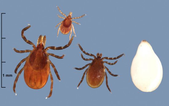
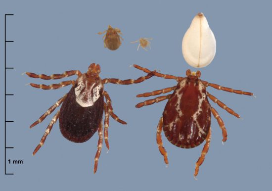
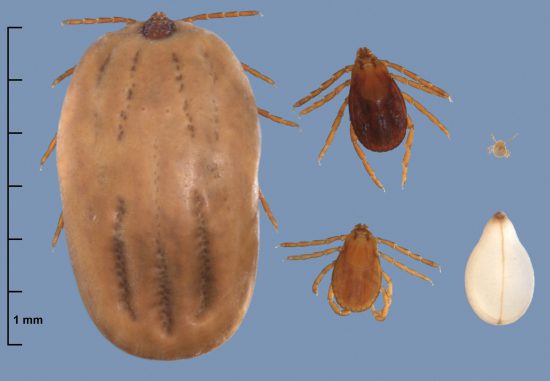
*All tick photos courtesy of Kent Loeffler, Department of Plant Pathology and Plant-Microbe Biology, Cornell University
