The Diagnostic Challenge
Definitive diagnosis of amelanotic melanocytic tumors is often difficult; these tumors can closely mimic poorly differentiated malignant neoplasms such as carcinomas, soft tissue sarcomas, and round cell tumors. However, it is crucial to accurately diagnose melanocytic tumors, as prognosis and therapy vary greatly between the differentials. Immunohistochemistry (IHC) is often required to confirm the diagnosis.
The Prognostic Challenge
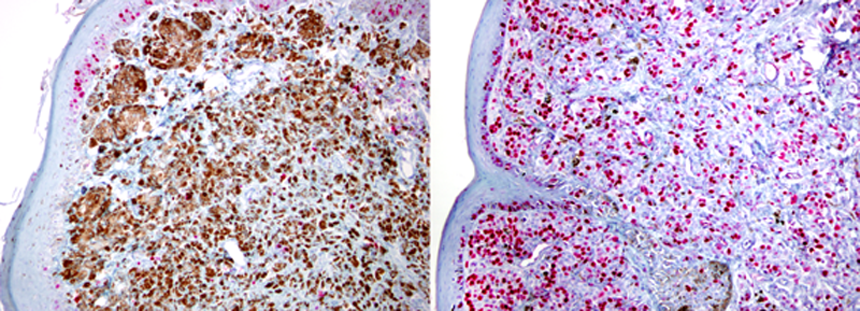
Accurately determining the prognosis for canine melanocytic tumors, especially those of the oral cavity, is difficult. Recent evidence suggests that a subset of canine oral melanocytic tumors may have a more favorable prognosis than historically thought. Nuclear atypia is a predictive histological feature for oral and cutaneous melanocytic tumors; however, it is subject to inter-observer variation and is difficult to assess in some tumors. Mitotic count is also helpful in predicting prognosis, but is not as predictive as nuclear atypia or Ki-67 index. The nuclear protein Ki-67 (MIB-1) is expressed in all proliferating cells and can be detected by IHC. The Ki67 index has been shown to be highly predictive of prognosis, based on survival times, for both oral and cutaneous melanocytic tumors..
Diagnostic and Prognostic Panels Offered
We offer a diagnostic melanocytic tumor panel that includes IHC labeling using a cocktail of antibodies against Melan-A, PNL-2, TRP-1, and TRP-2. This IHC cocktail is highly sensitive and specific in detecting amelanotic melanocytic tumors and is efficient and cost-effective.
A prognostic melanocytic panel includes immunolabeling for Ki67, and assessment of nuclear atypia, mitotic count, and degree of pigmentation for prognostic information.
Sample Submissions
All tests can be performed on routine formalin-fixed biopsy material as well as previously submitted biopsy samples. Alternatively, clients can request to have another pathology service submit the paraffin block or unstained slides to our laboratory if the tissue was originally processed elsewhere. For more information, please contact the Anatomic & Surgical Pathology laboratory at 517.353.1683, or see our catalog of available tests.
