
The Diagnostic Challenge
Differentiating IBD from enteropathy-associated T-cell lymphoma (EATL) type II (small cell) in cats is extremely difficult. Both conditions are most commonly diagnosed in middle aged to older cats of any breed and sex. The most common clinical signs with both diseases include vomiting, diarrhea, weight loss and changes in appetite. In addition, food hypersensitivity, parasitism, and endocrine, renal, or hepatic disease can also cause similar symptoms. Physical, ultrasonographic, endoscopic and microscopic examinations are of limited use in differentiating IBD from EATL in many cases.
Our Diagnostic Capabilities
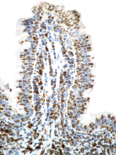
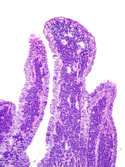
A step-wise diagnostic algorithm that first uses histomorphologic assessment, followed by immunophenotyping, and then PCR for Antigen Receptor Rearrangements (PARR) testing to determine clonality of lymphoid cells was developed in our laboratory to more accurately differentiate between IBD and EATL. Microscopic evaluation of routine formalin-fixed biopsy samples (endoscopic or full thickness) is used to screen for morphologic hallmarks of lymphoma, including marked lymphocytic infiltration of the tunica muscularis (full thickness samples only) and epitheliotropism (nests or plaques of lymphocytes within the epithelial layer). IHC is then used to differentiate between B- and T-cells, as well as to better visualize the location of T-lymphocytes in the epithelial layer and to identify heterogeneous inflammatory cell populations versus homogeneous neoplastic cell infiltrates.
The final diagnosis is based on the combined interpretation of morphology, immunophenotyping, and PCR tests for T- and/or B-lymphocyte clonality to differentiate neoplastic and inflammatory lymphocytes. At the MSU VDL we perform triplicate amplification, heteroduplex analysis, and capillary electrophoresis on a regular basis for diagnostic cases, which allows us to achieve the highest possible sensitivity and to avoid pseudoclones, which are false positive results. PARR results should never be interpreted independent of morphology. We therefore only offer PARR in combination with a biopsy or a second opinion and immunophenotyping.
Sample Submissions
All tests can be performed on routine formalin-fixed biopsy material as well as previously submitted biopsy samples. Alternatively, clients can request another pathology service to submit the paraffin block if the tissue was originally processed elsewhere. For more information, please contact the Anatomic & Surgical Pathology laboratory at 517.353.1683, or see our catalog of available tests.
