The following information outlines how cutaneous masses are routinely evaluated. To determine how other tissues are routinely evaluated, please see Submitting Special Tissues.
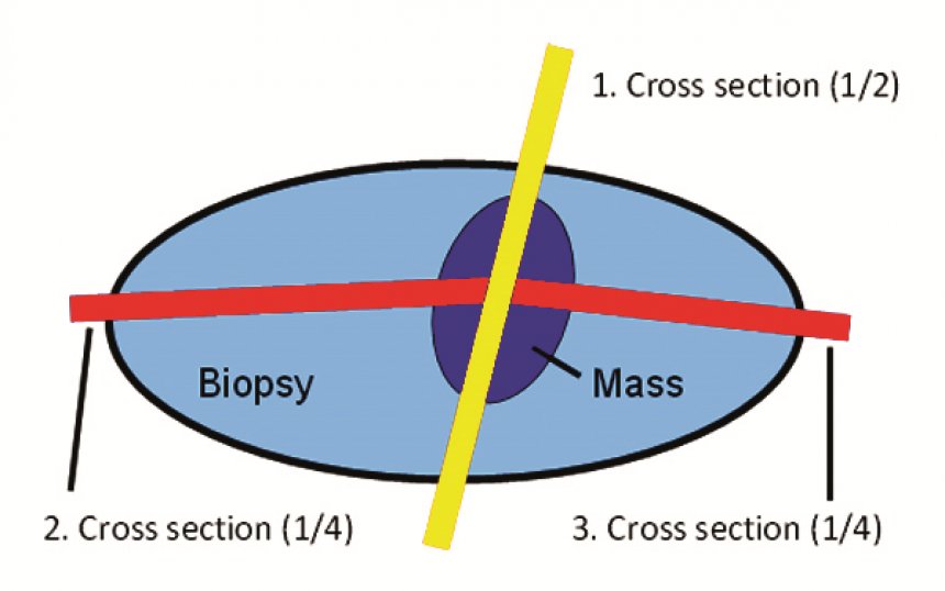
The standard trimming method used for routine sections is the “Cross Method” (i.e. ½’s and ¼’s):
- We palpate the specimen to determine where the mass/lesion comes closest to the surgical margins.
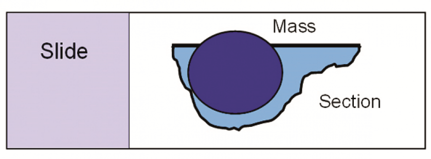
- Half-section [1/2]:
- We bisect the specimen through the mass/lesion so the section extends through the center of the mass.
- We then trim a 2-6 mm full thickness plane/slab/piece from this cut surface (this piece should demonstrate a cross section of the mass and the associated margins, see figures 1 and 2a).
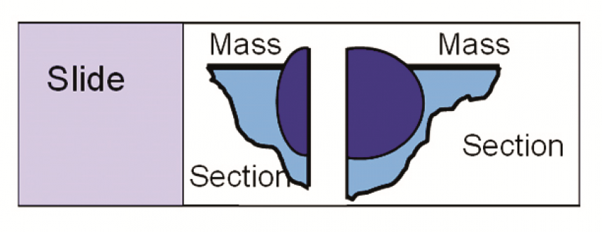
- Quarter sections [1/4’s]:
- We bisect each/both subsequent specimens (halves of the mass that have resulted from the cross section) through the mass/lesion along the longest axis of the tissue.
- Finally, we trim a 2-6 mm full thickness plane/slab/piece from this cut surface. These pieces should demonstrate the mass in a different plane, and the association of the mass with surrounding normal long axis tissue margins (when/where available), see figures 1 and 2b.
Figure 1: Dorsal view

Figure 2a: Lateral view of half section

Figure 2b: Lateral view of 2 quarter sections

Inked Margin Evaluation
- Evaluation of additional margins will only be performed following written or oral request.
- There are 3 options for how a specimen may be submitted for margin evaluation:
- Specimen is inked on all margins – see margin evaluation for process details
- Specimen has not been inked – we will ink specimen and proceed as above
- Some margins have been inked – we process only inked margins
- We always evaluate the quality of the ink and re-ink if the original ink is fading.
