The Challenges of Ocular Disease Diagnostics
As the organ of vision, the eye is highly specialized and wonderfully complex in form and function. Ocular disease can not only result in impaired vision or blindness, but often causes significant pain and distress. Reliable diagnosis of diseases affecting the eye is paramount for proper treatment of local disease, assessment of systemic disease state, and determination of prognosis with respect to either one or both eyes and overall patient health.
Many diseases that affect the eye and periorbital tissues are unique to these sites. Conversely, lesions in the eye may reflect generalized diseases. Diseases that affect one part of the eye often cause changes throughout the globe or in periorbital tissues resulting in complicated lesions that are often not uniform throughout diagnostic specimens. Given the complexity of ocular anatomy and pathology, accurate diagnosis of ocular disease requires specialized diagnostic expertise, careful attention to gross and histologic changes, and clear communication between pathologists and submitting veterinarians and ophthalmologists.
What Ocular Specific Expertise Can We Offer?
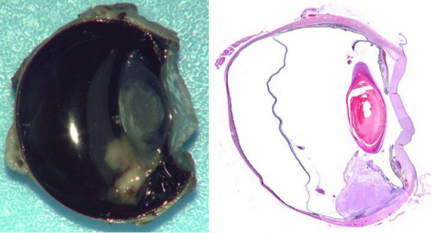
A veterinary pathologist with ocular pathologic expertise histologically reviews all ocular pathology specimens and provides detailed histopathologic descriptions of lesions along with interpretations of findings. Evisceration, enucleation, and exenteration specimens are grossly assessed and digitally imaged by pathologists which provides submitting veterinarians with an inside view of specimens during tissue processing. Other ocular biopsy specimens including conjunctival or corneal biopsies and third eyelid resections are also specially processed to ensure thorough evaluation including assessment of margins where applicable.
The MSU VDL’s full-service testing capabilities supplement quality gross and histologic evaluation of ophthalmic specimens. Such testing includes polymerase chain reaction (PCR), electron microscropy, immunohistochemistry, and in situ hybridization. In addition, the comparative ocular pathology service at our laboratory also seeks to fulfill research and educational needs through collaboration and making available digital gross images, detailed histopathologic descriptions, and histologic slides (additional fees apply to slides only).
Please contact the Anatomic Pathology Section at 517.353.1683, visit our catalog of available tests, and/or view the resources below.
For more information, see "The Challenges of Ocular Disease Diagnostics" in the Spring 2015 issue of Diagnostic News