Why the Eye?
Being the organ of vision, the eye and associated periocular tissues are highly specialized, wonderfully complex in form and function, and sensitive to disease. Ocular disease can not only result in impaired vision or blindness, but often causes significant pain and distress. Many diseases that affect the eye and periorbital tissues are unique to these sites, and can affect one or both eyes. Conversely, lesions in the eye may also reflect generalized disease or have wide-reaching health implications. Such is the case with hypertensive retinopathy, diabetic ocular changes including cataract and retinal vascular lesions, systemic bacterial and fungal infections, and neoplasms that either have metastasized to the eye or that have the potential to infiltrate into periocular tissues or metastasize from the eye to distant sites. Given these facts, assessment of the eye can provide valuable information regarding health specific to the eyes and to the overall health of animals.
Reliable diagnosis of diseases affecting the eye is paramount for proper treatment of local disease, assessment of systemic disease state, and determination of prognosis with respect to either one or both eyes, and overall patient health. Given the complexity of ocular anatomy and pathology, accurate diagnosis of ocular disease requires specialized diagnostic expertise, careful attention to gross and histologic changes, and clear communication between pathologists and submitting veterinarians and ophthalmologists.
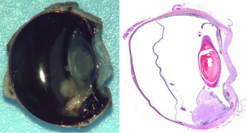
The eye is composed of many diverse tissue types derived from complex embryologic development. As such, different tissues within the eye react uniquely to insults, and because they have different embryologic origins, can also have distinctive congenital abnormalities. Specific common congenital abnormalities include corneal dermoids, goniodysgenesis or more extensive dysgenesis of the anterior segment, retinal folds, iridal or retinal colobomas, congenital cataracts, and micropththalmia. While each part of the eye can react differently to disease, diseases that affect one part of the eye often cause changes throughout the globe or in periorbital tissues resulting in complicated lesions that are often not uniform throughout diagnostic specimens. For example, even small benign iridociliary tumors are often associated with glaucoma due to the production of angiogenic factors by the tumors that when released into the closed system of the globe, result in the growth of fibrovascular membranes along the surface of the iris that can obstruct the filtration apparatus.
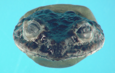
While many diseases of the eye cause extreme gross and histologic changes, other diseases such as degenerative retinopathies produce only subtle changes. Recognition of such subtle changes requires intricate knowledge of normal gross and histologic anatomy, clear communication of clinical presentation, and knowledge of eye specific pathologic processes. Diagnostic evaluation of ocular disease in the veterinary field is further complicated by differences in anatomy and function of eyes between the diverse species we encounter. Morphology and function of eyes and associated tissues can vary widely between species, as can the ocular diseases affecting the wide range of species we evaluate. For example, frogs have relatively simple eyes and when in captivity some species develop lipid keratopathy far more often than other animals. Birds such as owls and raptors have highly specialized and complex eyes that have rigid shapes due to the presence of bone within the sclera and commonly present with traumatic injuries.
What Ocular-Specific Expertise Can MSU DCPAH Offer?
The Michigan State University Diagnostic Center for Population and Animal Health (DCPAH) is committed to expanding and refining our comparative ocular diagnostic service. While we have already been providing excellent ocular pathology services to a small number of clients, we are excited to offer this expertise to a wider range of clients and to continue to foster innovative diagnostic testing and improved communication between pathologists and our clients.
This commitment to diagnostic ocular pathology begins with how specimens are submitted. Clear communication of clinical findings to DCPAH diagnosticians can be the difference between reaching a correct or an incorrect diagnosis. This is particularly true for globes submitted for histopathologic examination, as internal lesions may not be appreciable on external gross examination of formalin fixed specimens and may be missed unless direction is given by the submitting clinician. To improve communication of clinical findings to our diagnosticians, DCPAH has introduced a new ocular pathology submission form. This new submission form is designed to allow for easy and complete description of clinical ophthalmic findings along with providing areas for diagraming of ocular lesions to ensure that affected areas are specifically sampled.
Clarity in communication of diagnostic findings back to clinicians is as important as communication of clinical findings to diagnosticians. Ensuring clarity begins with how samples are processed upon receipt at DCPAH. For histopathologic examination, evisceration, enucleation, and exenteration specimens are always grossly assessed and digitally imaged by an ocular pathology specialist. Other ocular biopsy specimens including conjunctival or corneal biopsies and third eyelid resections are also specially processed to ensure thorough evaluation including assessment of margins, where applicable. Access to all digital gross images taken of specimens is provided through the DCPAH website allowing clinicians an inside look at diagnostic specimens and how samples are processed for histologic examination.
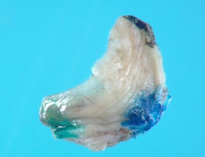
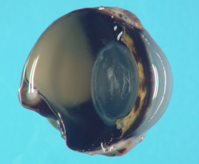
Given the challenges of ocular pathology diagnostics and the importance in clarity between clinicians and diagnosticians, the quality of histologic descriptions matters. Detailed histologic descriptions can explain clinically observed small changes that may not otherwise be reflected by only a bottom line diagnosis. In addition to interpretation of findings, every ocular pathology case receives a detailed description of all primary and ancillary findings, which is written or reviewed by our diagnostic ocular pathology expert to provide a complete, but concise, account of all histopathologic findings. The large number of pathologists at DCPAH, each with varied interests and expertise, provides great opportunity for consultation. Secondary review of ocular biopsies by experts in fields such as neoplasia, laboratory animal pathology, zoo and wildlife pathology, avian diseases, dermatopathology, and infectious disease is common and adds to the overall quality of ocular disease reports.
Not every case can be diagnosed with histopathologic assessment alone. Immunohistochemisty (IHC) is the most common ancillary tool employed in ocular pathology diagnostics. Any of the extensive breadth of immunohistochemical tests offered at DCPAH that are routinely used for tumor and infectious disease diagnostics can be applied to the ocular biopsies including our melanoma diagnostic panel. Specific for ocular diseases, we have validated multiple immunohistochemical tests targeting specific portions of the eye including ones that label specific parts of the retina and lens protein. PARR testing is also available to assess clonality of ocular lymphomas in formalin fixed specimens. For infectious diseases, DCPAH offers a wide range of in situ hybridization and PCR in addition to IHC that can be applied to formalin fixed tissues including both in situ and PCR for papillomaviruses and herpesviruses among others. As a full service diagnostic laboratory, DCPAH offers testing in 10 service sections outside of anatomic pathology. There is always the potential to submit fresh tissues, blood, or serum samples for tests like bacterial culture, PCR, clinical pathology, and endocrine profiles in addition to fixed ocular biopsy specimens. Going forward we will work to develop novel ocular disease-related tests and continuously expand the breath of our diagnostic tools.
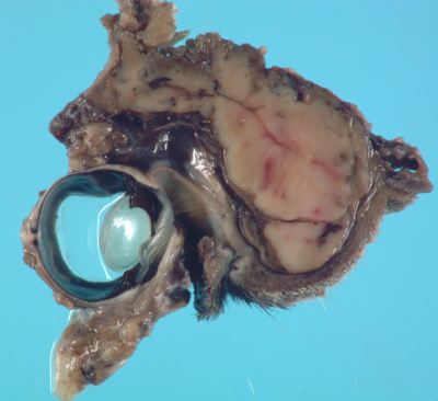
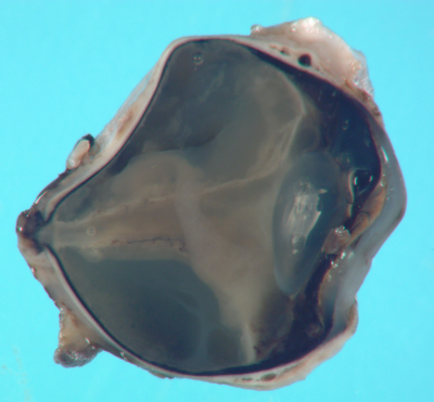
Beyond diagnostics, DCPAH also seeks to fulfill comparative eye-related research needs through collaboration and is committed to education. The full battery of histopathologic assessments, gross and histologic digital imaging, and molecular testing offered is available for collaborative research projects. Digital gross images of submitted specimens along with detailed histopathologic descriptions and recuts of histologic slides, which are available for additional fees, can serve as excellent educational materials for clients, students, and residents. In addition, diagnosticians are always available for consultation with referring veterinarians and specialists regarding specific cases.
