
Diagnosis of liver disease in dogs is aided by histopathologic evaluation of the liver, a crucial but often underused tool. Clinicians and pathologists working together via a team-based approach is recommended to ensure an accurate diagnosis and optimum patient management outcomes. Clinicians are encouraged to call the pathologist for case consultation prior to sample submission and/or after receiving the report. These discussions are often more helpful than simply interpreting the history from a submittal form or a biopsy report.
The Michigan State University Veterinary Diagnostic Laboratory offers a canine liver biopsy panel to increase diagnostic accuracy and interpretation of challenging canine liver cases. This panel has been modified slightly from our previous panel and has a new price effective 1/1/2026. The panel now automatically includes:
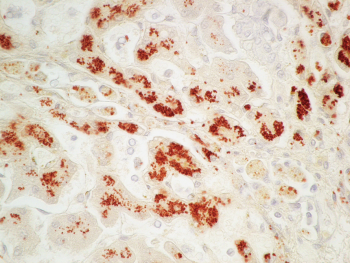
- mineral analysis on all cases (performed on fresh or formalin-fixed liver specimens)
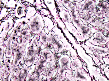
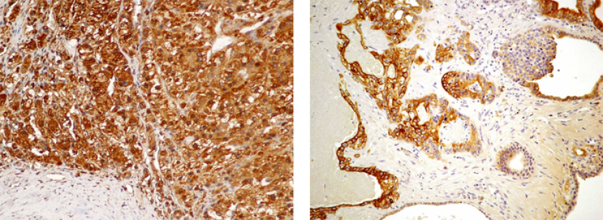
- up to four specialized histochemical stains, immunohistochemistry for CK19 (biliary epithelial marker)
- a scored/graded evaluation of type and degree of inflammation/degeneration, fibrosis/nodularity, and location and degree of copper accumulation when indicated
- and, in most cases, consultation with a pathologist specializing in liver diseases
Additional charges will apply for additional immunohistochemical markers and/or bacterial culture. If bacterial culture or other testing is desired on submitted fresh liver specimens, that testing must be clearly indicated on the submission form, so the fresh sample is not automatically submitted for mineral analysis. We recommend submitting two separate fresh specimens when both mineral analysis and culture are desired. Additional immunohistochemical markers are not included in the canine liver biopsy panel but may be recommended by the pathologist after reviewing the histopathology, as needed.
If mineral analysis cannot be performed for a particular case (e.g. liver specimens too small, only neoplastic tissue is present, other), the price of mineral analysis will be deducted from the panel fee.
The canine liver biopsy panel can be performed on any canine liver biopsy but is especially recommended for dogs with elevated liver values of unknown cause, suspected copper associated hepatitis cases, and suspected or atypical vascular anomaly cases. A standard biopsy may be sufficient for animals with hepatic neoplasms; however, pathologists may still recommend special stains to rule out concurrent copper associated hepatopathy or more accurately assess lobular architecture, or immunohistochemical markers to characterize the neoplasm. This can be done on an a la carte basis.
For cats and other species, we recommend ordering a standard biopsy at this time. The pathologist will make recommendations if any additional tests are thought to be beneficial for those cases and those tests will be available for additional fees upon request by the client.
For canine liver biopsy panels, if a fresh liver specimen was submitted, that specimen will be submitted for mineral analysis. If a fresh liver specimen was not submitted, after histologic review, any remaining formalin-fixed liver specimens or the formalin-fixed paraffin-embedded liver specimens will be submitted for mineral analysis, provided they are large enough. The biopsy and results of any additional testing will then be reviewed by a pathologist that specializes in liver diseases in most cases. All of these are included in the price of a canine liver biopsy panel.
If the pathologist recommends additional immunohistochemical labeling in the biopsy report, the client will need to call the Veterinary Diagnostic Laboratory to add that testing for an additional fee. Immunohistochemical labeling is especially helpful for vascular anomaly cases, including ductal plate malformations, and for neoplasms.
Lymphoma panel testing (immunohistochemical labeling and PCR tests for clonality), PCR tests for infectious organisms, and cultures and sensitivities are NOT included in this panel.

Liver biopsies can be challenging cases to review and often involve more time and discussions, often in conjunction with our veterinary internists. Thus, providing a complete and detailed history, including signalment, clinical signs, clinicopathologic findings, imaging results, gross findings, results of any other tests, and locations of the biopsy(ies) is extremely helpful.
For more information on collecting samples for a liver biopsy submission, please see our Recommendations for Liver Sampling.