Interpretation of liver biopsies can be challenging and often involves additional time and discussion among pathologists as well as communication with the submitting clinician. In order to provide our clients with the most accurate interpretation of these cases, additional testing such as evaluation of special stains, immunohistochemical markers, and/or heavy metal analysis, is often required. Therefore, to better serve our clients, we now offer a Premium liver biopsy panel (test code 40090) which will be available in our test catalog Thursday, September 1.
The premium liver biopsy panel includes histologic evaluation of the biopsy by at least one pathologist who specializes in liver disease who will automatically order specialized stains and/or immunohistochemical markers and/or heavy metal analysis as needed for an accurate diagnosis. This panel can be performed on any liver biopsy, but is especially recommended for challenging internal medicine cases, suspected vascular anomaly cases, intrahepatic neoplasms, and suspected cases of copper-associated hepatitis (CAH), including any liver biopsy from a dog breed that is prone to CAH (e.g. Bedlington Terriers, West Highland White Terriers, Labrador Retrievers, Doberman Pinschers, Dalmatians, etc.).


How Does It Work?
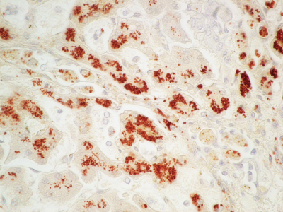
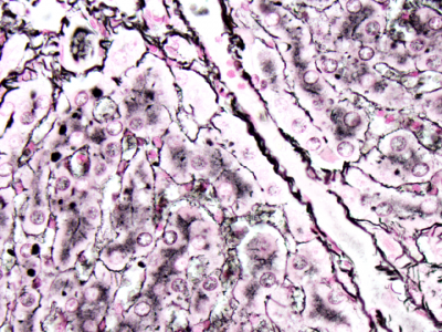
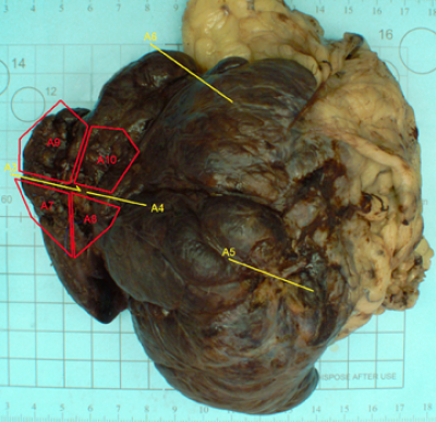
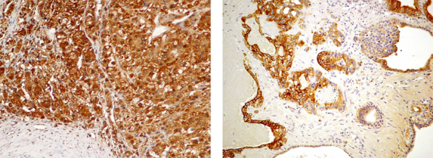
The liver biopsy is first reviewed histologically by one or more liver pathology specialists who then decides if up to three special stains (Trichrome for fibrosis, Rhodanine for copper, and Reticulin for lobular architecture) or up to two immunohistochemical markers (for vascular a n o m a li e s o r neoplasms), and/ or heavy metal analysis (quantitative evaluation for copper, iron, lead, selenium, and zinc) are required. When whole lobes with intrahepatic neoplasms are submitted, the surgical margins will be evaluated and sectioning of these margins will be documented via images that are available on our website. These tests are included in the price of the panel and are ordered automatically if enough tissue is available and if deemed necessary by the pathologist. This Premium liver biopsy panel can be performed on any standard formalin-fixed liver biopsy sample. If an infectious cause is suspected, submission of a fresh frozen sample of liver is also strongly recommended. If needed, bacteriology and/or virology testing can be performed on fresh samples for additional fees. Additional immunohistochemistry and insitu hybridization testing is also available for other infectious agents.
When requesting this panel we require a complete and detailed history that includes signalment, clinical signs, clinicopathologic findings, imaging results, gross findings, and results of any other tests. Ideally, multiple liver biopsies that represent multiple liver lobes should be submitted for every case. For certain disease processes, such as vascular anomalies, the histologic features can vary greatly between liver lobes and some lobes may appear normal histologically. Thus, vascular anomalies can be missed if only a single liver biopsy is submitted. Other diseases, such as copper associated hepatitis, can be masked by secondary chronic changes such as fibrosis and nodular regeneration. Submission of multiple biopsies can increase the likelihood of identifying less chronically affected areas that still contain large amounts of copper.

Samples from different lobes, or those representing different gross appearances in the same lobe, should be submitted in separate properly labeled containers or otherwise identified in order to ensure the most accurate evaluation. If the samples are small, such as those obtained via needle biopsy, samples from different lobes can be submitted in separate tissue cassettes or rubber free tubes such as microcentrifuge tubes filled with formalin and properly labeled. Larger samples, such as whole lobes, could be identified with different colors or numbers of sutures or different colors of ink.
Noting the location of samples and gross findings on the submission form is extremely helpful to the pathologist. Please also indicate if samples are taken from nodular lesions or any other masses. For example, if the pathologist is not aware that a sample is taken from the center of a hyperplastic nodule, and there are no bordering normal hepatocytes in the sample, a misdiagnosis of steroid hepatopathy may be made, as hyperplastic nodules often contain glycogen-laden hepatocytes.

Why Test for Minerals?
In recent years, we have diagnosed increased numbers of cases of canine CAH, especially in Labrador Retrievers, but we have also seen sporadic cases of CAH in a number of different dog breeds as well. Suspected CAH has also been reported in the literature in small numbers of cats and in two ferrets. In addition, we have found that liver copper values have been increasing in dogs since the 1980s. A combination of environmental and genetic effects is likely responsible for this increase.
To our knowledge, the DCPAH toxicology laboratory is the only veterinary diagnostic laboratory that offers validated quantitative analysis of hepatic copper, iron, lead, selenium, and zinc in formalin-fixed paraffin embedded tissues. Thus, we can accurately determine the amount of these elements in small liver samples after they have been completely embedded and reviewed histologically. This allows the pathologist to directly correlate what is seen histologically in Rhodanine (copper stain) stained sections with a quantitative value from the same section, as opposed to a separate liver sample that may exhibit different findings. Quantitative analysis can even often be performed on needle biopsy samples if several samples are taken and combined. Ideally, the total amount of liver submitted for element testing should weigh more than 50mg.
For more information on price, specimens, collection protocol, shipping requirements, and other additional information, please see our catalog of available tests or call us at (517) 353-1683.
