
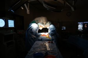
The Interventional Radiology Service at the MSU Veterinary Medical Center uses advanced imaging techniques, including ultrasound, fluoroscopy, CT, and MRI to guide the delivery of materials for diagnostic and therapeutic purposes. This minimally invasive approach to treatment facilitates shorter hospital stays, decreased pain, and a more rapid return to function. Our service is one of the few formal Interventional Radiology services in the country.
Patients typically visit the Interventional Radiology Service for:
- Respiratory interventions
- Cardiovascular interventions
- Urogenital interventions
- Portosystemic shunt treatments
- Oncologic applications
- Nutritional applications
The Interventional Radiology Service does not have specific service hours. However, we are always available during business hours for consultation with referring veterinarians to discuss how Interventional Radiology can aid in the treatment of many diseases of many small-animal patients. If we are unavailable, consultation calls will be returned promptly. The Interventional Radiology Service operates on an appointment basis; however, we recognize that certain disease processes necessitate emergency treatment, and we are able to meet these needs.
Respiratory Interventions
- Tracheal collapse has traditionally been treated surgically through the placement of prosthetic tracheal rings around the trachea to support it from the outside. We offer tracheal stent placement for dogs with tracheal collapse. Tracheal stent placement avoids surgery and is associated with a far lower acute complication rate than standard surgical treatment. The procedure is painless and animal normally return home the following day. Dogs with tracheal collapse will be assessed using physical examination findings and fluoroscopy (real-time x-ray). Ongoing medical management, prosthetic ring placement, and tracheal stent placement all have their place in the management of this challenging disease process. The best treatment recommendations will be made for your pet based on the extent and location of collapse.
- Tracheal Tumors / Strictures: Traditionally treated through surgical resection, tracheal stent placement offers a non-surgical option for palliation of clinical signs associated with these conditions.
- Tracheal or Bronchial Foreign Body Retrieval may be performed using fluoroscopic guidance.
- Chronic Pleural Effusion (accumulation of fluid around the lungs): Chronic pleural effusion is a life-limiting condition encountered due to chylothorax and various types of cancer. IR techniques are utilized to place indwelling drains in a non-surgical fashion linked to subcutaneous access ports allowing clients and their veterinarians to remove this fluid on a chronic basis without the need for painful repeated thoracocentesis (use of a needle or catheter to remove fluid from the chest).
Cardiovascular Interventions
- Vascular foreign body retrieval. The most common vascular foreign body is an IV catheter that has broken off or is severed and is lodged in the pulmonary vasculature. Using fluoroscopic guidance, vascular foreign bodies can be retrieved without the need for surgery.
- Pericardial Drain Placement: Pericardial drains are often placed on an emergency basis in animals with cardiac tumors that cause bleeding into the sac around the heart. The drain allows for the removal of this blood allowing cardiac function to return to normal. The drain is most often used as a bridge to surgical resection of these tumors.
- Balloon Pericardiotomy: Balloon pericardiotomy (creating a hole in the pericardium using a balloon under fluoroscopic guidance) is a minimally invasive approach to the long-term management of pericardial effusion (accumulation of fluid in the sac around the heart) due to certain types of cancer and idiopathic causes. Balloon pericardiotomy is inferior to pericardectomy (surgical removal of the pericardium) and thoracoscopic pericardial window (removing a “window” of the pericardium to allow fluid to drain), however, it is much less invasive and can result in sustained improvement in clinical signs.
- Diagnostic Angiography Studies: The IR service performs a variety of angiographic studies for the diagnosis and subsequent treatment of a variety of vascular anomalies including developmental anomalies, peripheral venous malformations, arterio-venous malformations, thromboembolic diseases (blood clots), vascular occlusions due to tumors, etc.
- Vascular Stenting and Angioplasty: Angioplasty and vascular stenting may be indicated for obstructed blood vessels due to tumors, clots, or other abnormalities. The IR Service is able to perform diagnostic studies and then therapeutic procedures to alleviate or palliate these conditions.
- The MSU Cardiology Service is equipped for the diagnosis and treatment of a variety of developmental cardiac diseases of dogs and cats in a minimally invasive fashion (balloon valvuloplasty, cardiac catheterization).
Urogenital Interventions
- Urethral Stent Placement: Dogs (and rarely cats) develop cancer that can cause partial obstruction of the bladder or the urethra making it difficult for them to urinate. Traditional treatment focused on placement of an indwelling tube into the bladder (cystostomy) via a surgical approach. Placement of a urethral stent across the narrowed region can restore urination in these patients without the need for any surgical procedure, the complications (chronic infections) that accompany it or the challenges that owners have to endure to intermittently drain the urine from the cystostomy tube. Placement of the urethral stent treats the clinical sign of difficulty urinating. The IR Service and Oncology Service work closely on these cases. The Oncology Service will develop a long-term treatment plan and present you with options for specific treatment of the tumor itself.
- Percutaneous Nephrostomy Tube Placement: A percutaneous nephrostomy tube is a tube that is used to divert urine from the kidney to the outside when the ureter (located between the kidney and bladder) is obstructed. Ureteral obstruction most often results from stones, infection, and tumors of the bladder. The procedure is performed under a short anesthesia and eliminates the need for emergency surgery in these often critically ill patients until a time when they are more stable and better surgical candidates.
- Ureteral Stent Placement: Ureteral stents are placed using a combination of IR and endoscopy techniques to allow urine to pass between the kidney and the bladder when an obstruction is present that is not amenable to surgery or when the client wishes to attempt to avoid surgical intervention. These procedures are performed with Dr. John Kruger who is an internist with extensive experience and expertise in endourology.
- Intra-arterial chemotherapy: Intra-arterial chemotherapy is designed to deliver large doses of chemotherapy directly into the major blood vessels feeding a given tumor. Bladder, urethra, prostate, and intrapelvic tumors of other origin may be amenable to this treatment approach. Please see Oncology Applications below for additional applications of IR procedures in the treatment of tumors.
- Antegrade Urethral Access: Antegrade urethral access is a technique that is utilized to facilitate placement of a catheter in the bladder when routine placement techniques fail due to the size of the patient, the presence of a tumor, or from a tear in the urethra. This technique is very quick and simple to perform and allows the clinicians of the IR Service to avoid surgical interventions that would normally be necessary to resolve many of these problems (surgical cystostomy tube placement, perineal urethrostomy for urethral tear or surgical placement of a urinary catheter).
Portosystemic Shunt Treatment
- Background: Dogs of all sizes are occasionally born with an abnormal communication of blood vessels in the liver (intrahepatic) or outside the liver (extrahepatic) that diverts blood flow around, rather than through the liver. The clinical signs resulting from this developmental problem include failure to grow and thrive, lethargy, depression, and abnormal behaviors including but not limited to blindness, tremors, seizures, star gazing, head pressing, and ataxia (being off balance). Extrahepatic shunts are most common in small breed dogs (especially Pugs and Yorkshire Terriers). Surgery is most often indicated in these patients and is highly successful. Intrahepatic shunts (IHPSS) are most common in larger breed dogs. Breed dispositions include the Labrador Retriever, Flat-Coat Retriever, Irish Wolfhound, and Bernese Mountain Dog, but all breeds may be affected (including the rare small breed). Traditional treatment of intrahepatic shunt has also been surgical using a variety of different techniques and devices. However, the shunt is often difficult to identify within the liver, challenging to dissect, and challenging to partially occlude. Frequently a second surgical procedure is necessary later in the life when the process must be repeated. The procedures have also been associated with unacceptably high complication rates including death (7-66% perioperative death rates have been reported). An endovascular (inside the vessel) repair technique has been developed and perfected that eliminates the need for open surgery, eliminates patient pain and discomfort associated with surgery, shortens hospitalization, and is associated with a very low perioperative mortality rate <5%). MSU has never lost a patient in the perioperative period. Instead of opening the abdomen and part of the chest to make the repair, the entire procedure is performed using catheters, stents, and coils from within the blood vessels themselves. The entire procedure is performed via a <5mm incision over the jugular vein in the neck. The procedure is called a Percutaneous Transjugular Coil Embolization (PTCE).
- Percutaneous Transjugular Coil Embolization (PTCE): MSU VMC is one of only three centers in the country offering this revolutionary treatment. A true team approach to treatment is needed for success. We work closely with the Anesthesia team during the PTCE procedure. The procedure is technically demanding and requires a familiarity with a variety of different techniques, equipment, and materials. Dr. Beal has performed more of these procedures successfully than any other veterinarian in the world with the exception of the Team of Drs. Weisse and Berent at the University of Pennsylvania with whom he did his training. If you or your veterinarian believes that your dog may have IHPSS, please call Drs. Beal and Mehler for consultation. We will help your veterinarian in the medical stabilization of the condition through medication and dietary modification. Once stable, your dog will be admitted to the hospital and a CT scan performed to identify the exact anatomy of the shunt. The PTCE procedure is performed days to a few weeks in the future. The CT and PTCE cannot be performed on the same day because of the contrast agents utilized and the risks that a high dose of contrast in a single procedure could provide.
Oncologic Applications
- Transarterial Chemoembolization (TACE): For non-resectable liver tumors.
- Transarterial Chemotherapy or Embolization: For palliative treatment of unresectable neoplastic disease. Example: Embolization for pain control for metastatic or primary bone tumors. Potential delivery of chemotherapy as a radiation sensitizer.
- Stenting of malignant urethral or ureteral obstruction (see above)
- Stenting of malignant vascular obstruction
- Stenting of malignant airway obstruction when surgery is contraindicated or not desired.
- Stenting of malignant gastrointestinal obstruction when surgery is contraindicated or not desired.
- Subcutaneous Vascular Access Port (SVAP) placement
Nutritional Applications
- Nasojejunal (NJ) feeding tube placement using IR techniques. Placement of the NJ tube allows for enteral nutritional support in animals with critical illness and vomiting or regurgitation. Feeding directly into the gastrointestinal system has been conclusively shown to be superior to parenteral routes of nutritional support (Total Parenteral Nutrition (TPN))
- Percutaneous gastrostomy tube placement
- Percutaneous gastrojejunostomy tube placement
- Esophagojejunostomy tube placement
Miscellaneous Procedures
- Glue embolization of hepatic arteriovenous malformations
- Glue embolization of thoracic duct
- Biliary drainages
- Esophageal stricture stenting (in concert with balloon dilation to decrease necessity for repeated dilations).
- Nasal embolization for intractable epistaxis. Unlike carotid artery ligation, this procedure may be repeated as needed.
- Repair of complex vascular malformations
- Subcutaneous Vascular Access Port (SVAP) placement
- Miscellaneous drainages
- Indwelling drainage catheters with subcutaneous access port
- Diagnostic peripheral angiography
Introduction
Following the trends in human medicine, there is an ongoing effort to adapt and develop minimally invasive therapeutics for the management of various problems facing veterinary patients. Minimally invasive therapeutics offer the advantages of smaller incisions, decreased pain, shortened anesthesia times, and shorter length-of-stay compared to traditional open surgical approaches. Currently in veterinary medicine, laparoscopy, thoracoscopy, minimally invasive orthopedic procedures, endourology, and interventional radiology are meeting this demand.
Interventional radiology (IR) involves the use of contemporary imaging modalities, such as fluoroscopy, endoscopy, ultrasound, CT, and MRI (or combinations thereof), to gain access to different structures in order to deliver materials for therapeutic purposes. IR is a subspecialty of radiology in human medicine. IR techniques have been widely utilized in human medicine for the past 20-30 years to effect minimally invasive diagnostic and therapeutic outcomes. Applications of IR in veterinary medicine are just being realized. The purpose of these proceedings is to present current applications of IR in veterinary medicine.
Equipment and Training
Many IR procedures require advanced imaging modalities. Fluoroscopy is a critical tool for performing most IR procedures. In IR, an array of guide wires with various properties, catheters specifically adapted for individual procedures and anatomy, stents composed of different materials and configurations, embolic coils, embolic particles, drainage devices, surgical glue, oils, chemotherapeutic agents, occlusion devices, balloons, etc. replace the standard surgical pack.
Because IR is so new to veterinary medicine, there are only two formal training programs in veterinary interventional radiology. They are at Michigan State University and the Veterinary Hospital of the University of Pennsylvania. It should be noted that many of the already developed veterinary IR procedures could result in significant harm or even death if improperly performed.
IR Applications in Veterinary Medicine
Potential applications of IR techniques for the management of veterinary disease states are boundless. The following paragraphs describe four common IR procedures involving the respiratory, hepatic, urogenital, and gastrointestinal systems. The table at the end of the proceedings describes other common IR applications being performed in veterinary medicine.
Tracheal collapse is a common affliction of small breed dogs. Traditional management of tracheal collapse is centered on medical management (cough suppressants, corticosteroids, management of concurrent problems). Surgical management using prosthetic rings placed around the trachea is an option in patients with cervical tracheal collapse that fail medical management. Surgical management of tracheal collapse tends to be invasive and is associated with a significant incidence of complications including but not limited to disruption of the tracheal blood supply, injury to the recurrent laryngeal nerve causing laryngeal paralysis, and the inability to access the intrathoracic trachea.
Tracheal stenting involves the placement of an intraluminal self-expanding metallic stent that holds the trachea open. Placement requires the use of fluoroscopy. Tracheal stenting offers a very rapid, non-surgical (everything is done through the airway) treatment option for animals with tracheal collapse. The incidence of acute complications is very low when compared to prosthetic ring placement provided the stent is sized and deployed appropriately. Long-term complications may include inflammatory tissue formation at the ends of the tracheal stent. This problem tends to be very steroid responsive and resolves rapidly. Stent fracture is another complication and occurs primarily in animals that continue to cough severely. As a result, ongoing medical management is still important although most patients require a less rigorous medication protocol.
Currently, the author recommends tracheal stent placement for dogs with tracheal collapse that are not having good quality of life in the face of medical management, those with intrathoracic tracheal collapse, and those whose owner does not wish to pursue a surgical treatment option. Dogs with intrathoracic tracheal collapse and mainstem bronchial collapse often benefit significantly from tracheal stent placement although they often continue to cough due to the mainstem bronchial collapse. Cervical tracheal collapse may be treated through traditional surgical techniques or placement of a tracheal stent. The entire procedure takes approximately 30 minutes and usually requires only 24 hours total hospitalization. It is important for clients to recognize that tracheal collapse is a progressive condition. This being said, tracheal stent placement offers an excellent palliative treatment option.
Malignant urethral obstruction: Transitional cell carcinoma, prostatic carcinoma, and other intrapelvic neoplasia may result in urethral obstruction. Traditional therapy has been centered on diverting urine via surgical placement of a cystostomy tube while pursuing traditional tumor-directed therapies. Cystostomy tube placement requires surgery and requires significant owner maintenance for the duration of the pet’s life. In addition, complications including tube dislodgement and recurrent, frequently multi-drug resistant urinary tract infection are not uncommon. Using IR techniques, an intraluminal self-expanding metallic urethral stent can be placed (non-surgically) via the vulva or penis to open the urethral lumen. Note that stents for this purpose are very different than those used for other applications. Using fluoroscopy, the length and width of the obstruction can be very precisely measured and a stent of an appropriate length and width to span the obstruction chosen. The stent is deployed from a delivery system introduced via the urethral orifice. The entire procedure takes approximately 1hour and is associated with little to no patient discomfort. Most often, patients are able to urinate immediately after stent placement. The greatest complication of the procedure is incontinence. Incontinence results from the stent spanning the urethral sphincter and at times, a significant portion of the urethra. The overall incidence of incontinence after stent placement is <20%. Females logically have greater problems with this than males. In a case series by Weisse et al. and based on the MSU experience, no patients died in the short or long term due to recurrent urethral obstruction.1 With the symptom of the neoplastic condition palliated, chemotherapy, radiation therapy or other adjunctive treatments may be utilized to address the underlying neoplasia.
Intrahepatic portosystemic shunt: The literature clearly illustrates that dogs with intrahepatic portosystemic shunt (IHPSS) have a very high incidence of surgical morbidity and mortality (10-50%). A technique for the repair of these shunts via an endovascular approach has been perfected at the University of Pennsylvania and is being performed regularly at Michigan State University and 1-2 other centers around the country. The technique is called a Percutaneous Transjugular Coil Embolization procedure. In short, the exact anatomy of the shunt is identified using Helical CT Portography (CT Angiography). Once the anatomy of the shunt is identified, the repair is performed under fluoroscopic guidance. Under general anesthesia, a large sheath introducer is placed in the jugular vein for the introduction of catheters, wires and stent. The shunt is localized in relation to the caudal vena cava angiographically. An appropriately sized caval stent is placed in the caudal vena cava at the mouth of the shunt. Through the stent, embolic coils are placed to partially attenuate the shunt while concurrently monitoring portal pressures (through the same catheter). The resulting increase in portal pressure results in increased hepatic portal flow, subsequent liver growth and development, and thus patient growth and development. The patient recovers from anesthesia and is discharged the following day. Procedural time is approximately 1.5 hrs and there is only a 4mm incision over the jugular vein. Acute morbidity and mortality are extremely rare. Approximately 30% of dogs undergoing this procedure require additional coils to be placed 3-12 months later to further attenuate the shunt and further increase hepatic flow.
Nasojejunal tube placement: Enteral nutritional support is associated with decreased length of stay, fewer infective complications, and significant cost savings when compared to parenteral nutritional support in people with critical illness. Nasogastric and nasoesophgeal tube placement is quick and the procedure is generally well-tolerated in small animal patients. However, many of these patients demonstrate nausea or vomiting associated with feeding into the stomach. Feeding distal to the stomach allows for the provision of enteral nutritional support in this patient population. Traditional surgical jejunostomy is invasive (requires surgery or laparoscopy) and is associated with significant complications including ostomy complications and septic peritonitis. A technique has been described for fluoroscopically-guided nasojejunal tube placement by Wohl et al, however, the technique was associated with a significant incidence of failure to achieve transpyloric passage and failure to achieve jejunal access. Tubes that only reached into the duodenum were frequently associated with oral migration. We describe a technique also utilizing fluoroscopy in which a combination of berenstein catheter and 260cm hydrophilic guidewire are utilized to gain guidewire access to the jejunum. Finally, the catheter is removed and a feeding tube is placed over the guidewire and sutured adjacent to the nasal planum. In our experience, ability to achieve transpyloric access is 100%. Ability to gain jejunal access is 84%. Oral migration is very rare. This technique has become standard of care in our service in patients with pancreatitis, septic peritonitis, and conditions associated with protracted vomiting or gastric motility disorders.
Additional Veterinary IR Procedures
Oncologic Applications
- Transarterial Chemoembolization (TACE): For non-resectable hepatic neoplasia.
- Transarterial Chemotherapy or Embolization: For palliative treatment of unresectable neoplastic disease. Example: Embolization for pain control for metastatic bone disease. Potential delivery of chemotherapy as a radiation sensitizer.
- Stenting of malignant urethral or ureteral obstruction
- Stenting of malignant vascular obstruction
- Stenting of malignant airway obstruction when surgery is contraindicated or not desired.
Stenting of malignant gastrointestinal obstruction when surgery is contraindicated or not desired.
Nutritional Applications
- Nasojejunal feeding tube placement using IR techniques.
- Percutaneous gastrostomy tube placement
- Percutaneous gastrojejunostomy tube placement
- Esophagojejunostomy tube placement
- Urinary applications
- Percutaneous nephrostomy tube placement
- Ureteral stent placement for urolithiasis
- Ureteral stent placement for malignant ureteral obstruction (bladder neck)
- Urethral disruption (iatrogenic or traumatic): Antegrade wire access with retrograde locking loop catheter placement into bladder. Indicated when retrograde catheterization fails. Frequently obviates the need for surgical intervention as the urethra heals over the catheter.
Miscellaneous Procedures
- Vascular foreign body retrieval
- Tracheobronchial foreign body retrieval
- Glue embolization of hepatic arteriovenous malformations
- Glue embolization of thoracic duct
- Biliary drainages
- Esophageal stricture stenting (in concert with balloon dilation to decrease necessity for repeated dilations).
- Nasal embolization for intractable epistaxis. Unlike carotid artery ligation, this procedure may be repeated as needed.
- Repair of complex vascular malformations
- Balloon pericardiotomy for recurrent pericardial effusion
- Pericardial drain placement for pericardial effusion decompression
- Miscellaneous drainages
- In-dwelling drainage catheters with subcutaneous access port
- Diagnostic peripheral angiography