Dr. Sara Jablonski is studying how to better diagnose and treat protein-losing enteropathy (PLE) in dogs. Both local and out-of-state clients may participate, as some studies require only 1 clinical visit. Clients whose dogs are part of 1 or more of Jablonski’s studies could save up to $2,500 on clinical services.

If you’re interested in applying for a PLE study with Jablonski, email the address linked below with information about your dog that will help determine their eligibility.

Please note: Jablonski and her collaborators are looking for dogs who have not seen prior treatment, and, most importantly, have not been treated with steroids.

Apply to Jablonski’s PLE studies.

About PLE and the Studies

What is protein-losing enteropathy (PLE)?

Protein-losing enteropathies are not a specific disease. PLE describes a condition—a syndrome—in which dogs lose an abnormal amount of protein from their gastrointestinal (GI) tract.

PLE is caused by GI disorders, the most common of which are intestinal lymphangiectasia (IL) and chronic inflammatory enteropathy (previously referred to as inflammatory bowel disease in dogs). However, PLE can result from neoplastic, infectious, mechanical, endocrine, and miscellaneous diseases and processes.

How common is PLE, and how dangerous is it?

According to insurance data, GI conditions are consistently among the top 3 reasons for veterinary visits. In addition, approximately 20 percent of dogs with chronic GI disease develop PLE.

While it isn’t the most common disease, PLE has a high mortality rate; veterinarians will typically inform owners that their dog with PLE has a 50 percent chance of survival.

How do veterinarians diagnose PLE?

To diagnose PLE, veterinarians must first exclude other causes of low serum protein. Then, a step-by-step process is followed to help determine the cause of the PLE. Understanding the cause will help dictate how the PLE is treated.

How do veterinarians currently treat PLE?

Because PLE can result from various GI diseases, there’s no cookie cutter approach. Veterinarians treat each dog as an individual. Therapeutic approaches include dietary changes, immunosuppression, and supportive care.

Why are studies necessary?

To treat PLE, veterinarians must obtain a lot of health information about each individual patient. These necessary diagnostics, as well as the treatments, are expensive for clients.

The gold-standard diagnostic workup includes small intestine biopsies that are obtained via endoscopy, a less invasive approach when compared to traditional surgery. This scope, in addition to the previous testing to exclude other causes of PLE not diagnosed with endoscopic biopsy, can cost clients between $4,000-5,000. Thus, studies are necessary to evaluate novel diagnostic approaches.

Treatment costs can vary, but add up quickly as part of a long-term care plan. Expenses for necessary prescription and/or homecooked diets can be significant, and some medications can be costly. Thus, new investigations demonstrating which treatments work best for individual cases of PLE are needed.

In addition, approximately half of dogs diagnosed with PLE do not respond well to treatment for reasons that are still unclear. More studies are necessary to help further understand of this syndrome and attempt novel therapeutic approaches to see if they may be more successful than traditional approaches.

Could PLE research in dogs translate to medical benefits for people?

Yes, there is a potentially translatable component of PLE research. People can develop PLE secondary to IL (Waldmann’s disease) or inflammatory bowel disease (such as Crohn’s disease). PLE research in dogs could help researchers address PLE in people (and vice-versa).

What are the advantages of participating in a clinical study?

Dogs that participate in clinical studies receive more attention from clinical teams, as well as treatments that may not otherwise be available. Dog owners can benefit from significantly defrayed costs of diagnostic and follow-up testing.

Img 3247 Edit
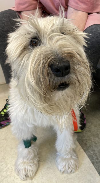
Tessa, a 5 year-old, female, spayed, soft-coated wheaten terrier with protein-losing enteropathy.
Figure4
Presence of small intestinal pinpoint to coalescing “white spots” consistent with dilated lacteals in a 5 year-old, female, spayed, soft-coated wheaten terrier with histologically diagnosed marked intestinal lymphangiectasia and the clinical syndrome of PLE.
Figure5
Photomicrograph of marked intestinal lymphangiectasia and moderate lymphoplasmacytic, neutrophilic, and eosinophilic duodenitis in a 5 year-old, female, spayed, soft-coated wheaten terrier with PLE. Photo credit: Victoria Watson, DVM, PhD, DACVP.