The doctors on our Orthopedic Services are highly trained specialists, performing surgery in five different state-of-the-art operating rooms, using the most advanced equipment and materials available today.

Interlocking Nailing
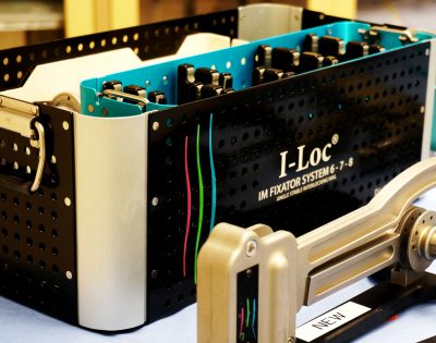
Interlocking nails are used as an alternative to bone plates in the treatment of long bone fractures. Interlocking nails are intramedullary rods locked to the bone via screws or bolts. Because of their size and position in the bone and because they can be applied remotely via small skin incisions, interlocking nails have mechanical and biological advantages compared to bone plates.
iLoc is a system Dr. Loic Dejardin patented and manufactured with BioMedtrix. Michigan State University currently holds the patent.
Facilities
Our patients are housed within the orthopedic ward in a private cage or run. We take patients out two to three times daily as their conditions permit. Depending on patient needs, we have access to and can consult with other specialists through the hospital.
Computer Assisted Pre-Operative Planning
While careful and accurate pre-operative planning is always important in orthopedics, it becomes critical when considering joint replacement, fracture repair particularly via minimally invasive surgery, and revision surgery. To assist our orthopedic surgeons, we have acquired highly sophisticated software that allows for 3-D reconstruction of bone structures from CT and/or digital radiography data. Dedicated templates of common implants including bone plates, interlocking nails and joint prostheses are used to accurately optimize size, shape and position of these implants for best possible outcome. Thanks to this high-tech, state-of-the-art approach, our orthopedic surgeons are able to considerably reduce intra- and post-operative morbidity while optimizing long-term clinical outcome.
Intra-Operative Image Intensification
Intra-operative image intensification is a technology that allows surgeons to view a moving image of the anatomic region of interest, using X-rays during surgery. This usually involves the use of a C-arm (or smaller, mini-C-arm) fluoroscope to acquire images while operating. The major advantages of this technology are 1) it allows orthopedic surgeons to safely place implants, such as rods, screws, and plates, under direct visualization, maximizing patient safety, and 2) it allows surgeons to operate using less invasive surgical techniques, which minimizes trauma to tissues and may help patients to heal faster after surgery. Intra-operative image intensification can be used in many procedures. One of the most common applications is fracture repair. A C-arm fluoroscope can be used to place pins in anatomic regions that present high risks of complications if the implants are not precisely placed, such as in the case of fractures of the spine. A C-arm fluoroscope can also be used to place pins to stabilize fractures using external fixators, to allow for reduction of the fracture and subsequent stabilization without having to make a large surgical incision. This can help to minimize recovery time and optimize function of the affected limb.
