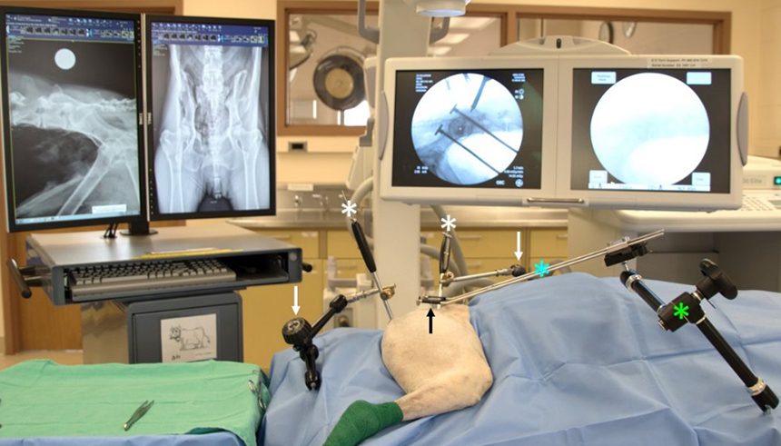
A novel Sacroiliac Luxation Instrument System (SILIS) was devised at Michigan State University to address the limitations associated with C-MIO repair of SIL/F. This photo shows the different components of the SILIS. The MIO reduction handles (white *) inserted in the ischiatic tuberosity and ilium are linked to table-bound 6-axis reduction arms (white arrows). A Minimally Invasive Lucent Aiming Device (MILAD, black arrow) is connected to a table-bound 6-axis reduction arm (green *) via a custom designed sliding handle (blue *).

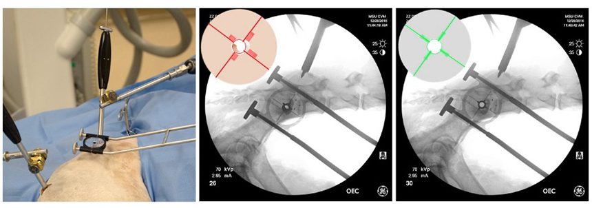
The MILAD is inserted through a stab incision made at the level of the sacral body (left). The MILAD crosshair and fins allow for rapid and accurate alignment over the safe sacral implantation corridor (center and right). The MILAD position is adjusted until accurate orientation (e.g., superimposition of the crosshair and the fins) is achieved (right).

Postoperative radiograph of a SIL/F treated with a lag screw applied via open reduction (ORIF) (left). Note the craniodorsal screw location in relation to an ideal position in the sacral body (crosshair). Postoperative 3D CT reconstruction of a bilateral SIL/F treated using MIO assisted with the SILIS-MILAD (N-MIO technique) (right). Both screws are located along the sacral body safe implantation corridor. Studies at MSU have demonstrated the superiority of MIO over ORIF in terms of screw orientation and location. The current study aims to evaluate clinical outcome and surgical safety.
