There is not much about Loki that is small. Not his size, not his energy, and certainly not his place in Hannah Rissman’s life.
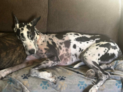
The six-year-old Great Dane loves walks, snuggles, and just about every dog he meets. His owner, Hannah Rissman, calls him her “Oafy Dofy Loafy” because more often than not, he is happiest simply lying around and being loved.
But after Loki was injured by a gunshot, Rissman didn’t know what the future would hold for her happy, gentle companion.
“It was a really scary situation,” Rissman says. “I didn’t know what the outcome would be.”
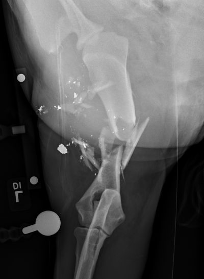
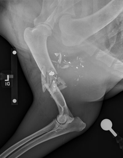
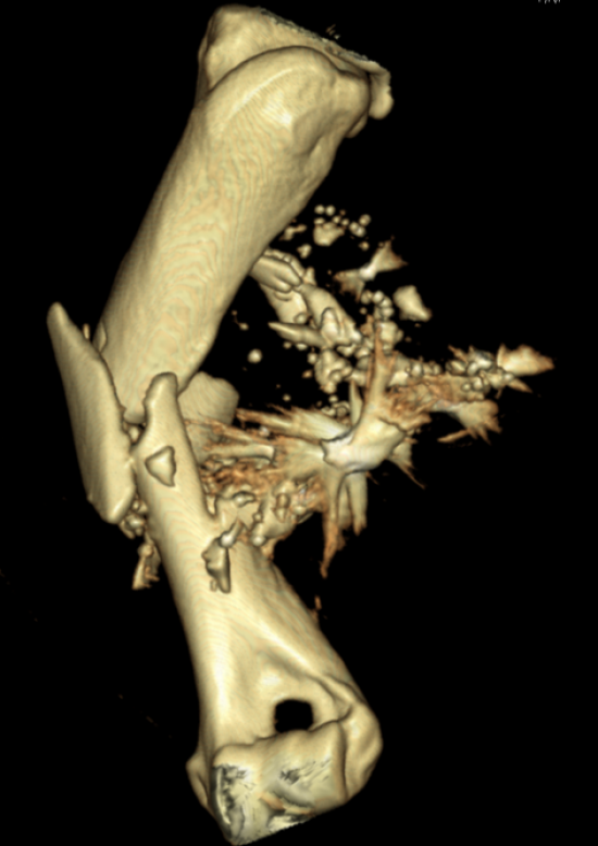
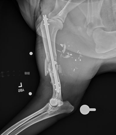
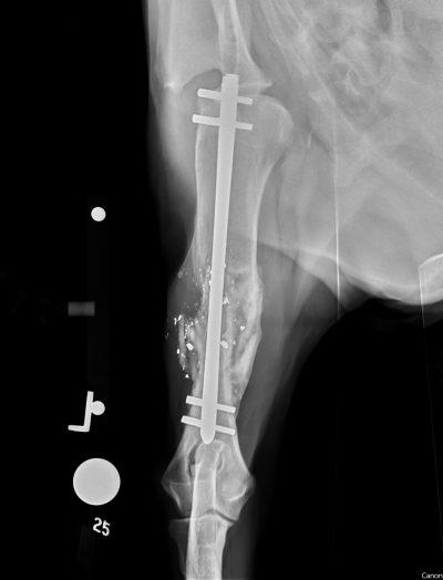
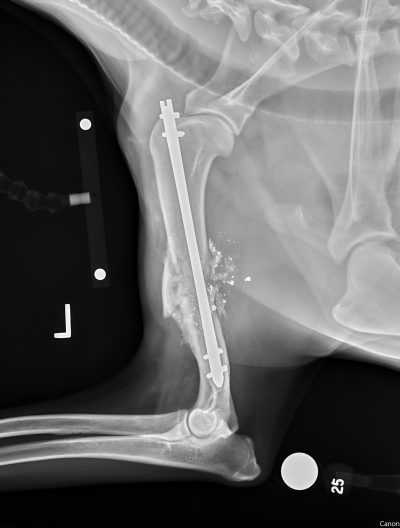
Rissman brought Loki to the Michigan State University Veterinary Medical Center, where he was assessed for a gunshot wound to his left front limb. The injury caused an open fracture of his left humerus—the large bone extending from the shoulder to the elbow—as well as significant damage to the surrounding muscles and skin.
After Loki was stabilized through the Hospital’s Emergency Service, he was evaluated by the Orthopedic Surgery Service to determine whether his leg could be saved. Because of the severity of the injury and the extensive soft-tissue trauma surrounding the fractured bone, the orthopedic surgery team determined that a minimally invasive approach was the best option.
“Cases that sustained a lot of soft-tissue trauma on top of the fracture itself are good candidates for minimally invasive approaches,” says Dr. Maria Podsiedlik, assistant professor of orthopedic surgery and fellow candidate in the Minimally Invasive Orthopedic Surgery Fellowship Program. “We don’t want to add more trauma by creating a big opening through the skin and dissecting the muscles to get to the bone.”
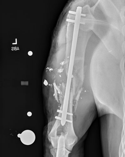
Instead of making a long incision to expose the entire fracture, the surgical team made two smaller incisions—one near the top of the humerus and one near the bottom—allowing them to repair the fracture without directly visualizing the broken bone. Using intraoperative fluoroscopy, a type of real-time X-ray imaging, the team guided an interlocking nail through the bone to stabilize the fracture.
That approach makes an important difference in cases like Loki’s. Traditional fracture repair can require surgeons to dissect through muscles and other soft tissues to reach the broken bone. With a minimally invasive approach, surgeons can preserve more of the tissues surrounding the fracture, including the fracture hematoma, which contains growth factors that support healing and a faster recovery.
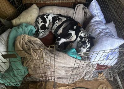
For Loki, the approach proved successful. When Podsiedlik saw him again two weeks after the surgery, he was walking well on the injured limb. The dramatic swelling that had accompanied his initial injury had subsided, and his leg was beginning to look normal again.
Keeping Loki quiet during his recovery, however, proved to be another challenge.
“Even when he first got home, he was breaking out of my back patio,” remembers Rissman. “He was busting onto my porch and running up the steps, even though he was barely supposed to be walking.”
Rissman laughs at the memory of trying to convince her energetic Great Dane that he had just been through a major injury.
“I was like, ‘Dog! Do you realize that you have been shot? You need to calm down.’”
Today, Loki is “practically back to normal,” Rissman says. She credits the team at MSU with getting Loki through a very scary and serious situation.
“Beyond the groundbreaking science at MSU, all of the doctors I worked with just had incredible bedside manner,” says Rissman. “I’m so grateful. I think if it weren’t for MSU, I would have a three-legged dog right now.”
At the Forefront of Orthopedic Surgery Worldwide
Loki’s case is a great example of the advanced orthopedic procedures available through the Orthopedic Surgery Service. Specifically, it showcases the expertise MSU offers as an official Minimally Invasive Orthopedic Surgery (MIS) Fellowship Training Center designated by the American College of Veterinary Surgeons (ACVS).
MSU is currently one of only four academic institutions worldwide offering this ACVS Fellowship training in small animal orthopedic minimally invasive surgery. The highly specialized program builds on the knowledge and experience surgeons gain during an ACVS surgery residency, preparing them to become recognized experts in a specific area of surgical practice.
For patients like Loki, that advanced training creates opportunities to consider specialized treatment approaches that may not be available at every veterinary hospital.
“This MIS Fellowship Program is beneficial because it allows us to provide an even higher level of expertise and to create standardized training for future minimally invasive orthopedic surgeons,” says Podsiedlik. “Through our additional training, we are able to offer these high-end orthopedic procedures to our patients.”
The Fellowship focuses on minimally invasive approaches to orthopedic trauma, such as fracture cases, as well as arthroscopy, which uses a small camera and specialized instruments to diagnose and treat conditions within joints such as the shoulder, elbow, and knee.
The program also strengthens education and training, giving surgery residents, veterinary students, and veterinary nursing students opportunities to observe advanced orthopedic procedures and learn about cutting-edge treatment options. At the same time, it expands the options available to referring veterinarians when patients need specialized orthopedic care.
“If a case is amenable to a minimally invasive approach, that is our first choice for treatment,” says Podsiedlik. “MSU is unique in the equipment it can provide for these procedures, as well as the range of orthopedic surgeries, imaging, and postoperative care we can offer.”
For Loki, the expertise available at MSU gave him the opportunity to return to the life that he loves. “Loki has the most amazing personality,” says Rissman. “Being able to watch him get back to normal so quickly feels like witnessing a miracle.”
ACVS Fellowship Training CenterImages of Loki's case