Jane Hoel is a first-year DVM student at the Michigan State University College of Veterinary Medicine. Below, she shares about the College’s DVM curriculum and offers a snapshot of her recent experience.
Part One: Preparing for the Lab
The College’s reinvented curriculum has a totally different structure from its legacy, or traditional, model. Classes are structured as deep-dives into specific body systems. For first-year DVM students, classes run in two parts. First, we watch a lecture online and review study materials. Then, we meet together for a lab based on the lecture and other materials for the lesson.
Recently, I completed an ultrasound lab during our digestive system class. In order to prepare for the lab, I watched a video of an abdominal ultrasound being performed on a dog. I also watched a narrated PowerPoint lecture on abdominal ultrasound in horses. The canine ultrasound video was helpful to watch; the ultrasound was performed while I listened to a description of what the video was showing. The instructor would occasionally freeze the screen and show labels on organs. The video was extensive, and it was a good resource to view prior to our lab.
The horse video was in the supplemental materials, and I found that lecture video very helpful, and was glad that I viewed it ahead of time. Cartoon images of organs were superimposed on a picture of a horse, and these were used to help us visualize where the organs are located. They also included labeled, still ultrasound pictures of the organs, which I found very helpful.
Part Two: the Lab
For the hands-on ultrasound lab, our goals were to:
- Identify the liver, spleen, gallbladder, kidneys, and small and large intestines of the dog with ultrasound;
- Describe probe placement to visualize each organ;
- Identify many of the same structures on the horse; and
- Deduce the problems that would ensue if colonic function was disturbed in horses.
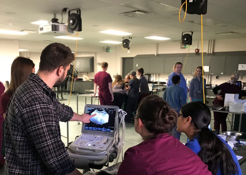
The first half of the lab, held in the LeBlanc Clinical Skills Lab, was learning how to do an abdominal ultrasound on dogs. We were in small groups of three or four students paired with a veterinarian and a veterinary technician. The veterinarian first ran through the structures we should see, quizzed us on our anatomy knowledge, and gave us tips for holding the probe. We each had a turn finding the diaphragm, liver, gallbladder, stomach, spleen, left and right kidneys, urinary bladder, and descending colon.

In the second half of the lab, we all headed to the Mary Anne McPhail Equine Performance Center for the horse ultrasound. We split into two groups to watch either the right or left side be imaged. On each side of the horse, a veterinarian ran through the abdominal structures we should be able to find on ultrasound. Each student conducted an ultrasound while the veterinarian asked us to find specific organs. On the right side of the horse, my group was able to find the cecum, right dorsal colon, and right kidney duodenum. We also found the lungs and the heart, even though they are not technically in the abdomen. On the left side of the horse, my group was able to find the stomach, spleen, large colon, left kidney, and the small intestines.
The Takeaway
My favorite part of the lab was being able to perform the ultrasound myself. I learn the best when I am able to perform tasks and skills, rather than just reading about them. Just moving the probe direction and angle could drastically change the image on the screen. By experiencing that myself, it was much easier to cement the concepts learned in the prep material. I really enjoyed this lab, and my fellow students agreed; many of us wished it could have lasted longer than two hours!
Through the reinvented curriculum’s combination of the prep material and the lab, we received valuable knowledge and experience for our future careers as veterinarians. The prep material helped me adequately prepare for the lab session. After watching the video lectures, I had an idea of what the abdominal organs looked like on ultrasound and how to describe the appearance of tissues on an ultrasound image (hyperechoic, hypoechoic, and anechoic).
In my opinion, the most valuable aspect of the reinvented curriculum is the exposure to extensive hands-on clinical skills and applications. It allows us to further cement ideas, concepts, or information learned in lectures and prep material. Some concepts, especially skills, can be hard to wrap your head around by just reading standard operating procedures. Having an instructor walk us through the steps and give us tips and tricks really helped me remember what I was doing and why I was doing it. We are not asked to just memorize anatomy and physiology; we are asked questions that apply our knowledge of normal anatomy and physiology to predict the potential clinical consequences if normal is disrupted. Since we are taught concepts using clinically relevant cases and skills, we are well on our way to graduate ready to begin our careers in veterinary medicine.


