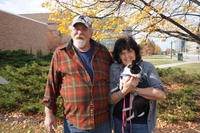
For Ed and Roseanna Launstein, few things matter as much as their pets. So when Pugsly, their then 8-month-old Boston Terrier, started vomiting up some food, seemed a little sluggish, and had less of an appetite, they took note right away. Two weeks later during her spay appointment, her veterinarian noticed that Pugsly had fluid accumulating in her abdomen. The veterinarian observed many unusual blood vessels and a strangely shaped liver.
The diagnosis

Pugsly was then referred to the MSU Veterinary Medical Center right away, where the clinicians observed the fluid accumulation and took note of the surgical findings. This, coupled with abnormal blood work (severely elevated bile acids), suggested a developmental problem involving the blood vessels, and thus function of the liver.
The clinicians had to find out what was causing this. A dual phase CT angiogram, a specialized CAT scan that gives excellent blood vessel detail, showed that Pugsly had multiple arteries from various locations (high-pressure vessels) feeding a nidus of small vessels (almost like the head of Medusa) and leaving through two large veins (low-pressure vessels). This is an extremely rare condition called hepatic arterio-venous malformation (HAVM). This condition was causing Pugsly to have high blood pressure in those veins draining the HAVM, resulting in her opening extra vessels called shunts to help disperse that pressure.
Pugsly’s clinical signs could have been managed with diet and medication, but there was no guarantee this would be effective over the long term. The other option was a surgical or image guided intervention to block off the veins draining the AVM. The Launsteins decided that an attempt to cure the problem was their best option.
“We wanted to give Pugsly a chance at a healthy life,” Roseanna said. “We love her so dearly.”
The surgery

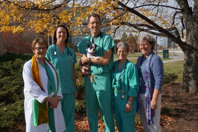
And so it was on. Pugsly would undergo a procedure that involved team members from various departments in the hospital: Interventional Radiology, Soft Tissue Surgery, Anesthesia, Cardiology, Diagnostic Imaging, and Critical Care Medicine. It was a risky procedure, and one that had only been performed once to the VMC’s knowledge.
But the detailed planning and expertise of the VMC team paid off. After a seven-hour procedure in which both traditional surgical techniques were melded with image guided minimally invasive techniques and vascular plugs within the draining veins of the AVM, Pugsly is recovering well and on the road to recovery.
“Pugsly is doing well, playing with her family,” Roseanna said, noting Pugsly’s playmate, a Papillion named Razzle Dazzle. “Dr. Beal is amazing, and we can’t thank him enough for giving Pugsly a second chance to have a more healthy and playful life. We are deeply in debt to him and the staff he chose. They were all just perfect.”
Looking ahead
The success of this surgery is big news for the veterinary world. The hospital hopes that Pugsly’s success will open doors for more successful outcomes for other dogs with this rare and debilitating problem in the future. Pugsly has another procedure set for March of 2016.


