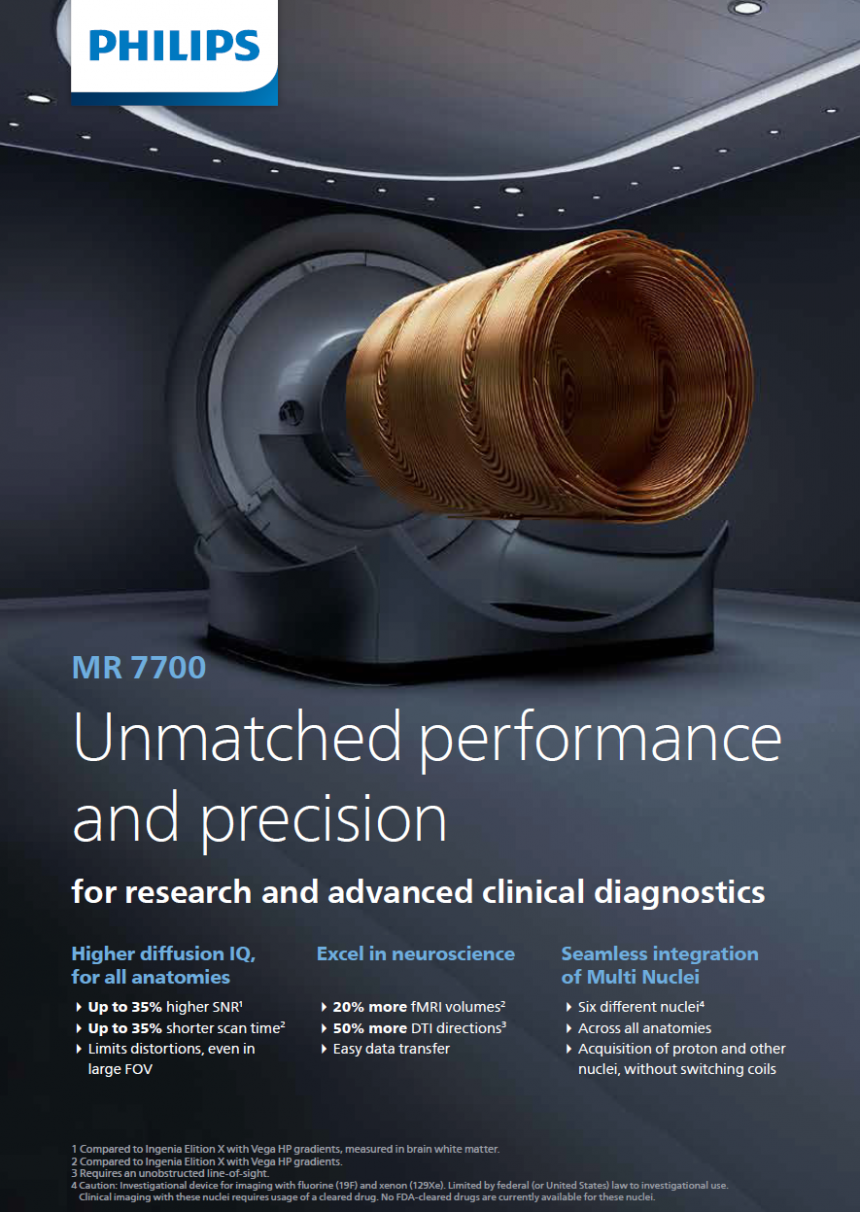
Highlights: MSU’s Philips MR 7700 3T MRI System
- Largest scannable field-of-view with overall shortest bore length
- High-performance gradients of 65mT/m at 200 T/m/s
- 32-channel head coil accessory
- Specialty-based software (neurological/neurovascular, cardiac, musculoskeletal, abdominal)
- More signal and resolution per voxel, and data points with fewer artifacts and delays
This time next year, the Philips MR 7700 3.0T MRI system with artificial intelligence and deep learning will be up and running at Michigan State’s Veterinary Medical Center. But the Hospital’s patients won’t be the only ones to benefit from the upgrade; researchers at MSU and beyond can reap the rewards of incredible image quality, faster.
“Together with Philips, MSU truly is pioneering clinical care and research capabilities in veterinary medicine,” says Dr. Srinand Sreevatsan, associate dean for the College of Veterinary Medicine’s Office of Research and Graduate Studies. “It will enhance the total activity and exposure of our College across campus and the state.”
The incoming 3.0T MRI will offer researchers and clinicians higher quality scans with reduced waiting time without compromising data, as well as the ability to image subjects of various sizes and species.
“A 3 Tesla MRI is so powerful, it gives great image quality with the ability to image everything from extremely small subjects like mice to the largest subjects like horses or wild cats,” says Rebecca Linton, manager of the Hospital’s Radiology Service. “And the new artificial intelligence software—the way it plans data points and understands what it’s processing—allows the MRI to enhance your image without compromising data integrity. We’ll get better image quality in less time, which is unheard of. It’s going to be beautiful.”

In addition to being at the cutting edge of imaging technology, the new Philips MRI system has a variety of software options that will help further neurovascular, cardiac, musculoskeletal, and abdominal research.
“It’s the newest, latest, and greatest in performance, and there’s specialized research software that can take scientists to new heights in their investigations. The new Philips MR 7700 3.0T is even capable of multi-nuclear imaging,” says Linton. “There are programs that can be written into grant costs, and that could enable or enhance a multi-million-dollar study.”
Get Ready to Research
Eager to make use of MSU’s new Philips MR 7700 3T MRI System? Contact Radiology Service Manager Rebecca Linton to discuss your project and set up a VetStar account.
The new MRI purchase comes amid the launch of MSU’s Clinical Innovations Program (CLIP), the College’s re-invigorated clinical trials initiative. The new imaging system’s benefits to CLIP are already apparent.
“Our new Philips MRI will be the cherry on top of veterinary clinical research here,” says Dr. Kelley Meyers, director of MSU’s Veterinary Medical Center. “The MR 7700 3T MRI opens a lot of opportunity for animal researchers, and that’s only complemented by our other services. As a hospital, we have the experts here to sedate and anesthetize research animals, as well as to care for and recover them.”
For more information about the Philips MRI coming to MSU, contact Rebecca Linton.
How Can I Prepare?
To get ready to use the new Philips MR 7700 3T MRI System at the MSU Veterinary Medical Center, researchers will need to provide:
- University account number
- Description of subjects, project timeline, and scan frequency
- Sedation/anesthesia needs
- Scan-building protocols and/or protocol builder contact information
- Point(s) of contact
