Teamwork to manage clinical trauma at the MSU College of Veterinary Medicine
A Collaborative Team
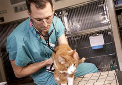
Doctors Matthew Beal, Ari Jutkowitz, and Amy Koenigshof have saved their share of lives, but the Emergency and Critical Care Medicine (ECCM) team has a broader mission: one that simultaneously advances education, research, and medical care for small animals.

“In addition to providing extraordinary clinical service, we are training the next generation of criticalists and developing new treatment modalities that let us save more lives every year,” says Beal, director of Interventional Radiology Services and chief of staff for the Small Animal Veterinary Medical Center.
Beal, Jutkowitz, and Koenigshof, diplomates of the American College of Veterinary Emergency and Critical Care, work closely with specialists across the College to provide a breadth of expertise and advanced technology unmatched in the state. The hospital works closely with the Departments of Small Animal Clinical Sciences, Large Animal Clinical Sciences, and Pathobiology and Diagnostic Investigation.
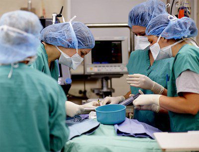
Many ECCM cases move through different services within the hospital. For instance, a critical trauma patient with multiple injuries may enter through the emergency room door, where he is stabilized before radiologists conduct diagnostic imaging. Next, the soft tissue surgery section conducts emergency surgery to repair a ruptured bladder. Once the most critical issues are attended to, he would then be transported back to the critical care team for on-going supportive measures before having his pelvis and femur fractures repaired by the orthopedic surgery service a day later. Specialists—and advanced technology—come together to treat patients that enter through the emergency room doors.
Advanced Care For Special Cases

A phone call from a Detroit-area emergency clinic to the MSU teaching hospital began the life-saving journey for Miley, a four-year-old Siberian husky.
Owner Ryan Vojtkofsky first took Miley to the Animal Emergency Center in Novi when she wouldn’t stop vomiting. Even with the administration of fluids, her condition worsened over the next two days. The veterinarian called MSU to review the case. “They said they thought they could help Miley,” says Vojtkofsky, “but if we were going to get treatment, we should take her immediately—that Miley’s chances of survival were dropping every minute.”
They suspected she had leptospirosis, a bacterial disease that can cause kidney and liver failure.
The drive from Detroit felt like an eternity for Vojtkofsky. By the time they arrived in East Lansing, Miley’s kidneys had stopped working. The Diagnostic Center for Population and Animal Health (DCPAH) quickly confirmed the suspected leptospirosis diagnosis.
Dialysis was Miley’s only hope. Artificially supporting kidney function can be done intermittently or continuously, but continuous therapy can be more successful in patients who are unstable or in critical condition. The emergency team began continuous renal replacement therapy (CRRT).
The CCRT equipment includes blood and fluid pumps, blood tubing, multiple filters, and a monitor that closely tracks blood flow rate, dialysis rate, ultrafiltration rate, and other parameters of dialysis. Success relies on the close collaboration of a team that is specially trained in CRRT. Concerned that a high-energy breed like Miley might become restless during therapy, doctors took extra precautions to ensure all of the tubes stayed in place over her two days on CRRT. Koenigshof played an important part. “On the first night that Miley was admitted, I stayed with her after she was put on dialysis. We had a little sleep over. I kept her calm and monitored her through the night.”
The morning after Miley arrived at the hospital, says Vojtkofsky, she exhibited phenomenal improvement, but treatment and recovery would keep Miley in the hospital for about two weeks. “One of the things that really helped me through the experience was how helpful and friendly everyone was,” he says. “I felt welcomed and was invited to stay as long as I wanted. The doctors called me with updates twice a day and on the third day, she was stable enough for me to visit,” he recalls.
For the two weeks she was in the hospital, he traveled from Novi every other day to visit. He walked Miley outside the hospital and spent time in the “quiet room,” which is situated away from the examination rooms. “It was so nice to be able to spend time with her like that.” After a few days on dialysis, Miley was weaned slowly from fluids and monitored to make sure her kidneys were functioning. After being discharged, Miley returned for a check-up two days later and then for a final check-up a week later. Now, Miley is back to her old self. Vojtkofsky describes her as playful and lovably hard-headed. “Everything is hers—or it will be,” he laughs. “And she is amazingly loving—she loves to snuggle.”
Research to Enhance Treatment
Along with expertise and technology, the rapid application of research findings into the clinical setting is saving lives every day at the VMC. Across the College, research projects are advancing knowledge in specialty areas of veterinary medicine, including but not limited to ophthalmology, oncology, cardiology, and interventional radiology—all of which may be important contributors to the treatment of patients who come in through the emergency room doors.

“In ECCM, our goal is to be able to provide the same level and availability of care to our animal patients as people might receive in a Level 1 trauma center or major university hospital,” says Beal. “We’re rapidly moving in that direction.”
The case of Vincent, a seven-year-old German shepherd, illustrates the capabilities of the ECCM service when integrated with the rest of the hospital.
Vincent arrived at the hospital after being hit by a snowmobile. He was in shock, had multiple femoral fractures, and a rapidly expanding hematoma in his left hind limb. The ECCM team applied counterpressure to stem the bleeding, and started fluids and transfusion with fresh frozen plasma and packed red blood cells, but they needed to find the source of the bleeding in order to stabilize the patient.
The anesthesia and interventional radiology teams joined the criticalists in order to identify the source, stop the bleeding, and preserve the local blood supply. The multi-disciplinary team was successful—they stabilized Vincent’s cardiovascular system and stemmed the blood loss. The orthopedic surgery team then repaired the fractures and the patient was returned to ECCM for ongoing support.
Translating Research Into Practice
Interventional radiology (IR), which helped stabilize Vincent, is a subspecialty of radiology in human medicine that can be used to diagnose or to treat patients. IR specialists use advanced imaging techniques including fluoroscopy (video xray), ultrasound, MRI, and CT to guide them as they thread tubes or small instruments through blood vessels and other pathways to reach the problem. Beal is one of the first veterinarians in the nation to receive formal IR training, and the hospital’s IR program is one of only four formal IR services in the country.
Procedures that typically require surgery may be treated in a minimally invasive fashion with IR techniques. One area of clinical and research focus of the ECCM and IR groups is the delivery of nutritional support to critically-ill patients. In animals that don’t tolerate nasogastric tube placement due to vomiting, traditional options include providing nutrition either intravenously or through a surgically-placed catheter that bypasses the stomach and delivers nutrition directly to the intestinal system. Research suggests that direct delivery to the small intestine is better than IV feeding, but the required surgery has a number of potentially fatal risks.
Beal and the IR team developed a number of minimally invasive techniques that pass a feeding tube from the patient’s nose or esophagus, past the stomach, all the way into the jejunum—the middle section of the small intestine. The procedure can be completed in just 20 to 30 minutes. “At the VMC, this is now our standard of care where the animal is intolerant of gastric feeding,” says Beal. His research on this technique was published in the April 2011 Journal of Veterinary Emergency and Critical Care.
The VMC launched the ECCM resident training program in 2005 with a single resident. Since then, five residents have completed residencies and all have become diplomates in the American College of Veterinary Emergency and Critical Care.
As of last year, more MSU DVM graduates were in resident training programs around the country than any other institution.
Clinical Patients Supporting Innovation
The collaborative services at the VMC provide exceptional patient care and unparalleled opportunities for teaching. They also provide tremendous discovery and research possibilities. In some cases, patients admitted to the VMC for treatment and therapy may be eligible to participate in a research project.
“The relationship between the hospital and the College benefits both animals and research,” says Dr. Elizabeth Ballegeer, assistant professor of diagnostic imaging. Ballegeer’s research, which has the potential to also contribute to human medicine, includes studies that correlate findings of non-invasive diagnostic imaging with other diagnostic methods. She recruits most of her study participants from the hospital.
“In many of our cases, dogs with a liver mass referred to the hospital for sonograms are able to receive more sophisticated and thorough imaging. Though we know of the mass that initially includes them in the study the PET/CT scan we conduct as part of research may reveal additional or distant lesions that were not detected in the sonogram,” says Ballegeer. “This allows the clinician to guide different diagnostics or surgical approaches that better address the patient’s whole disease, rather than just the single mass.”
A New Space for Specialized Care

The ECCM team treated almost 6,200 patients in 2012—a caseload more than twice what it was 10 years ago. As the service, team, and technology have expanded, however, the physical structure of the emergency care unit has remained unchanged. The College is now planning a renovation to the space that will support the VMC’s commitment to providing the finest care for veterinary patients, world-renowned veterinary education, and clinical research to advance care for animal and human health.
“Our expertise has grown, our technology has advanced, and our facilities need to be renovated to make the most of those improvements,” says Dean Christopher Brown.
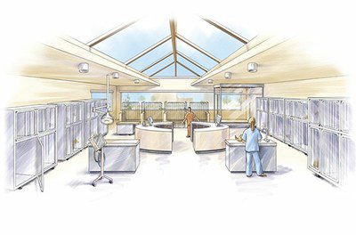
Currently, ECCM patients are housed in several areas, making close observation and response challenging. These several areas will be combined into one large open space that will optimize the continuum of care all within view of a centralized observation desk. The team will be able to maneuver mobile equipment—like the dialysis machine used on Miley—more easily. The staff will be able to respond more rapidly in crisis circumstances. Within view of caregivers, there will be an isolation ward with separate ventilation for patients with infectious diseases, respiratory, and/ or gastrointestinal problems.
As the number of patients continues to increase and the possibilities for veterinary medicine grow, the Veterinary Medical Center will be prepared to continue its leadership in the field of emergency and critical care medicine.
Posted: March 2013
Contact: Casey Williamson
