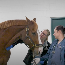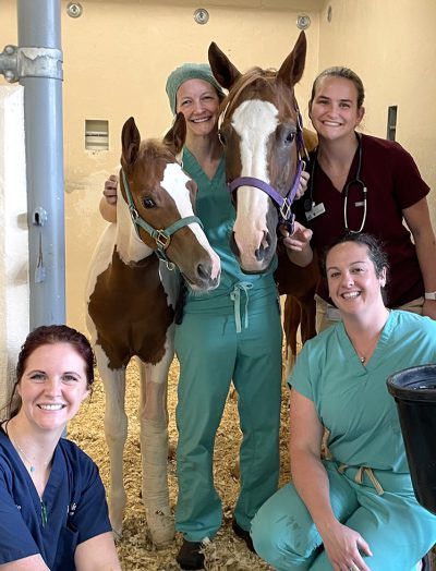
This story features faculty Dr. Ashley VanderBroek, and residents Dr. Jackie Willette and Dr. Lindsay Monroe.
Percy, a 3-month-old American Paint Horse colt, was presented to the Large Animal Emergency Service at the MSU Veterinary Medical Center with his mare for severe, acute left forelimb lameness.
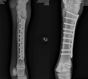
Radiographs revealed a closed, non-displaced, diaphyseal spiral fracture of the left third metacarpal (cannon) bone. Although the fracture was non-displaced at the time, it spanned nearly the entire bone and required stabilization to prevent the fracture from propagating further. Spartan veterinarians immediately began planning surgery for his fracture repair.
Surgical Procedure
Percy was stabilized the evening of presentation and was prepared for surgery the following morning. Based on the configuration of the fracture, a 9-hole, broad locking compression plate was applied to the dorsomedial aspect of the cannon bone. It was important that the plate spanned the entire diaphysis to appropriately stabilize the fracture and prevent forming a stress riser in the bone. Because Percy was a foal, it was also critical that the physis (growth plate) was not disturbed—as he had a lot of growing yet to do! Fluoroscopy was used throughout the entire procedure to assess the position and contour of the plate and also determine the appropriate length for each screw. Surgery went well and Percy did well under anesthesia.
Recovery and Outcome
Percy was placed in a full limb bandage with a caudal and lateral splint for recovery from anesthesia. This was done to protect the repair, as recovery from general anesthesia is not always graceful in horses. Just like in humans, animals recovering from anesthesia can experience dysphoria and ataxia. While dogs and cats can be confined to a cage or held until they are aware of their surroundings, horses’ recovery from anesthesia can be a critical event. Because they are prey animals, they will often try to stand before regaining their balance and may seriously injure themselves even in padded rooms with soft floors. Due to the colt’s small size, the team at MSU could hand-recover Percy and assist him once he was ready to stand. He ambulated back to his stall comfortably and was reunited with his mare. He was maintained on pre- and post-operative antimicrobials and anti-inflammatories.
The splints were removed the following day and Percy was transitioned to a distal limb bandage. He remained comfortable on the limb and had no difficulty ambulating around the stall. After several days of monitoring in the Hospital, Percy and his mare were discharged for continued stall rest at home. Recheck radiographs were taken at 3 and 8 weeks postoperatively and showed progressive healing of the fracture. Once fully healed, Percy was turned out in the pasture with his mare, where he could again run and play.
Commentary
“Fracture repair in horses has come a long way,” explains Dr. Ashley VanderBroek, assistant professor and surgeon with the Hospital’s Large Animal Service. “Even with advancing technologies and state-of-the-art facilities, there are many factors that put horses at a disadvantage when it comes to long bone fractures. One of these factors is their size. Although horses have very slender legs, the averaged sized adult horse weighs approximately 1,200 pounds. This puts a lot of weight on the recently repaired fracture and can result in implant failure if the construct is not strong enough. Unlike people that can be placed on bed rest for extended periods of time, horses need to be able to stand and walk immediately after surgery. More specifically, they need to be able to bear weight evenly on all limbs. The coffin bone of the horse is suspended within the hoof capsule via laminae. When a horse is unable to stand on one of its limbs, it overloads the other limbs causing the laminae to fail resulting in laminitis. This is an incredibly painful condition and unfortunately once support limb laminitis develops, there is not much that can be done.”
Another factor relating to equine anatomy that negatively impacts healing is that there is no muscle located below the carpus and tarsus—only bone, tendons, and ligaments. This means that there is minimal soft tissue covering the bone and many displaced fractures can easily become open and contaminated. Infection is a serious issue with fractures because it delays healing and causes lysis of the bone surrounding the implant. It is also incredibly difficult to resolve these infections without removing the implant. Thankfully, Percy’s fracture was non-displaced and closed, and he did not develop an infection. His small size and young age also greatly contributed to his success.
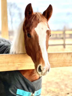
Thanks to his procedure, Percy returned to normal function. He was later able to successfully compete as a yearling in halter classes, and his owner reports that his movement seems unaffected by his injury.

