Presentation and Diagnosis
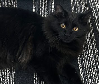
Duck was a four-month-old foster kitten when he presented to the MSU Veterinary Medical Center’s Cardiology Service for evaluation of a heart murmur.
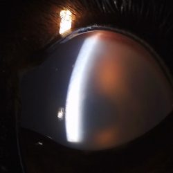
An echocardiogram (ultrasound of the heart) confirmed a grade V left basilar continuous murmur, a loud murmur often associated with a patent ductus arteriosus (PDA).
A PDA is a congenital heart defect in which the ductus arteriosus (the vascular communication between the aorta and pulmonary artery that exists during fetal development) does not close as expected at birth.
The ductus arteriosus is a normal fetal structure that allows blood to bypass the lungs before an animal (or human) is born. Usually, the ductus arteriosus closes at birth when the animal starts to breathe. However, in rare cases, the ductus arteriosus does not close, and a communication remains between the pulmonary artery, which takes blood from the heart to the lungs to pick up oxygen, and the aorta, which sends blood from the heart to the rest of the body.
When a PDA occurs, blood will flow out of the aorta and into the pulmonary artery. This abnormal flow of blood causes over-circulation of the lungs and the heart and can lead to left-sided heart enlargement and subsequent left-sided congestive heart failure. Failure to close or repair a PDA can result in extreme pressure changes, reversal of blood flow, de-oxygenation, and can result in death.
The echocardiogram, performed with Dr. Robert Sanders and Dr. Matthew Denton, revealed a left-to-right shunting PDA with moderate left atrial and left ventricular dilation.
Treatment
Duck was evaluated by the MSU Soft Tissue Surgery Service and plans to proceed with surgical ligation of the ductus arteriosus were made. Dr. Maureen Spinner, Dr. Kathryn Asatryan, and Dr. Meredith McClure performed Duck’s surgical repair.
Duck was placed in right lateral recumbency, and his left thorax was clipped aseptically scrubbed and draped in routinely. A standard approach to the left lateral thorax through the fourth intercostal space was made using a #10 scalpel blade, Metzenbaum scissor, and electrocautery for hemostasis. A Finochietto retractor was used to improve visualization.
Care was taken to avoid damage to the surrounding vagus and phrenic nerves. The surgery team isolated the abnormal vessel and applied special sutures to ligate and prevent blood flow through this structure.
Immediate cardiac auscultation confirmed that the continuous heart murmur had resolved upon placement of the surgical ligations. A thoracostomy (chest tube) was placed to remove the air that entered the thoracic cavity upon surgical entry. This thoracostomy tube was temporary and removed upon confirmation of the presence of negative pressure within the thoracic cavity. While anesthetized, Duck was concurrently neutered.
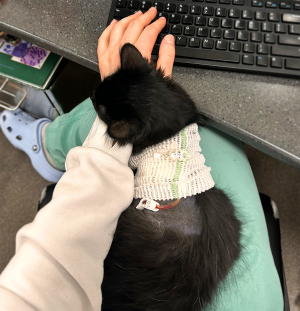
He recovered well from his procedure, and his prognosis following surgery was excellent. Duck has continued to thrive with his family, having been adopted by MSU rotating intern, Dr. Alex Garrett.