Initial Presentation and History
Claudia is a one-year-old mixed breed dog who transferred to the Capital Area Humane Society as a stray from an animal control organization. She was intact and had received preliminary vaccines from animal control.
Shortly after placement, Claudia’s foster noted bilateral eyelid swelling after spending extended time outdoors. Allergies were suspected, so diphenhydramine was initially prescribed, however over the course of a few days Claudia’s eyes became clouded and painful.
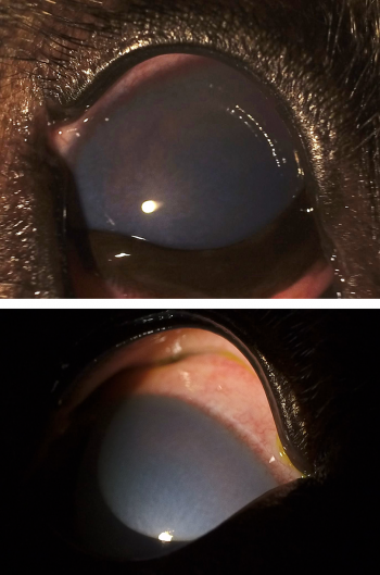
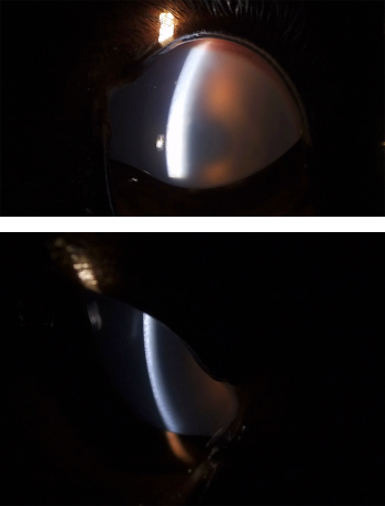
The shelter veterinary team examined Claudia and noted a “blue haze” that appeared to wax and wane throughout the day. Her left eye was particularly severe, with a cornea hazy enough to obscure the view into the iris. She was given oral pain medications.
Because of the cloudy appearance of the eyes, the shelter team suspected some sort of acute glaucoma in the left eye, possibly developing in the right. They procured a tonometer to measure intraocular pressure (IOP), with readings of 12, 16, 12, 11, 10 mmHg in the left eye and right eye readings of 20, 20, 21, 19, 22 mmHg. These readings were not consistent with glaucoma—dogs presenting with this condition usually have readings greater than 25, beyond 40 in severe cases, and even up to the 80s. Claudia was also unusually young for a dog with primary glaucoma. Secondary glaucoma was still possible. However, due to severe corneal clouding, they were unable to examine inside her eyes to investigate underlying ocular causes.
The CBC reported low normal RBC and mild elevation of white blood cells, neutrophilia, monocytosis, increased basophils. A fluorescein stain yielded negative uptake, and other lab work found no significant abnormality.
An ophthalmology team of board-certified clinicians, residents, and advanced clinical students from the MSU Veterinary Medical Center also visited the shelter to examine Claudia and provided a second opinion. The MSU team primarily noted severe corneal edema with possibly mild anterior uveitis. The IOPs measured at this visit were also within normal.
At this point, the top considerations for causes of Claudia’s corneal edema included immune-mediated, infectious, or degenerative diseases of the corneal endothelium. The corneal endothelium lines the inner surface of the cornea, and keeps the cornea dehydrated thereby keeping it clear. Blood was drawn for an infectious disease panel.
Diagnosis
A few days later, the infectious disease panel results arrived. The report showed a positive result for canine adenovirus type 1 (CAV-1), which causes infectious canine hepatitis. CAV-1 is associated with ocular lesions of corneal edema in about 20 percent of infected animals and in 0.4 percent of those that receive a modified-live strain of the virus in CAV-1 vaccines. In the latter cases, symptoms appear one to three weeks after inoculation.
However, these days, few animals receive the CAV-1 vaccine which is part of the DHLPP combination vaccines. Due to near-universal replacement with the DA2PP combination vaccines that contain canine adenovirus type 2 (CAV-2) immunizations which provides cross-protection against CAV-1. This makes “blue eye” rare, though the complication fit with Claudia’s presentation.
In digging through Claudia’s medical record, the shelter and MSU team found that Claudia had received a DHLPP (distemper-hepatitis-leptospirosis-parainfluenza-parvovirus) vaccine about three weeks prior, and investigations showed the vaccine she had received indeed targeted CAV-1.
Based on the timing of Claudia’s DHLPP administration, the CAV-1(+) on the PCR test, and her clinical signs, Dr. Keiko Miyadera (DVM, PhD, DACVO) of MSU made the diagnosis: “blue eye” associated with CAV-1 vaccination.
She explains:
“Clinically, Claudia’s condition did look primarily like endotheliitis, given the disproportionately severe corneal edema despite low-grade inflammation perceived in the anterior chamber of the eye. This was noticed by Dr. Melaney Mayes [veterinary ophthalmology resident at MSU] (also see her paper) and aligns with CAV-1’s tendency to target the endothelium.” The resultant edema or swelling of the corneal gives the eye a blue haze, hence the term “blue eye.”
As a young and healthy dog, Claudia’s prognosis was good with supportive anti-inflammatory medications. Unlike glaucoma, which likely would have resulted in enucleation, the CAV-1 vaccine reaction would clear up over time. Her eyes healed, and she was adopted a few weeks later.