By Andrzej Ogrodny, first-year resident for the MSU Internal Medicine Service
Featuring Daniel Langlois, DVM, DACVIM (SAIM), clinician for the Internal Medicine Service; Lauren Kustasz, DVM, third-year resident for the Internal Medicine Service; Olivia Lefere, MSU DVM Class of 2022; Jordan Guy, MSU DVM Class of 2022; Judy Eastman, LVT
History and Presentation
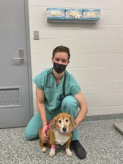
Riley is a 7-year-old female spayed mix breed dog who was presented to the Michigan State University Internal Medicine Service in December of 2021 for evaluation of recurrent bladder stones and for discussion of laser lithotripsy. Riley had a previous history of several urinary tract infections, and she also had one prior occurrence of bladder stones which were removed surgically. She was again diagnosed with a large, 2 cm. bladder stone this past summer (2021). When her bladder stone recurrence was noted, the owners attempted a medical dissolution protocol with diet and antibiotics, but the stone failed to dissolve. The owners hoped to avoid another repeat abdominal surgery; that is when a cystoscopy-assisted laser lithotripsy procedure was elected.
In February of 2022, Riley was represented for a drop-off cystoscopy with laser lithotripsy of the bladder stone. Previously, Riley had been managed for struvite stones with her regular veterinarian and had stones removed from her bladder in June of 2020. She has been on a urinary diet for the long term for management that she is reported to enjoy. At the time of presentation, she did not have any urinary signs (straining, posturing, or passing small amounts of urine). Riley continued to do well at home, and her owner noted that she is urinating normally with no lower urinary tract signs (no hematuria, or stranguria currently). Riley also has a normal energy level and appetite with no v/d/c/s. Her owner does not notice any urine leakage and Riley does not lick her vulva frequently.
Diagnosis

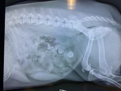
A single lateral radiograph was performed, which revealed a single radiolucent, spherical stone within the lumen of the bladder prior to her procedure. It measured 2 cm. x 2 cm. A urinalysis was performed by her primary care veterinarian prior to the procedure, which also was screened for any signs of a urinary tract infection.
Riley’s primary diagnosis was cystolith with historic struvite stones excised via her referring veterinarian. She also was diagnosed with obese BCS with a recessed vulva, likely secondary to body conformation. Her tertiary diagnosis was recurrent UTIs. This stone had not responded to previous empiric antibiotics and dissolution diets.
Prior to treatment, Riley’s urinalysis and culture had no evidence of bacterial growth (a typical screening before cystoscopy).
Treatment and Outcome
A venous blood gas, packed cell volume, and total solids were checked prior to anesthesia that were unremarkable.
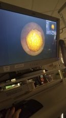
Riley’s procedure continued as planned; the cystoscope was introduced with ease. There was a normal vestibule opening, and the mucosa upon entry into the urethra was pink and smooth. On entry into the bladder, there was a large yellow-brown stone with a rough surface in the lumen. The caudodorsal aspect of the bladder had mild polyploid projections that were slightly hyperemic. Focal areas of hyperemia were noted within the bladder.
Laser ablation via YAG laser was then performed; the stone was hollow in the center and fragmented into multiple rough segments small enough to be snared. Pieces were retrieved with basket snares with mild amounts of bleeding and urethral inflammation sustained.
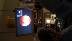
Urethral urohydropulsion was performed between each segment to remove segments of stone until urine voided was clear in nature. A visual assessment of the bladder after the procedures revealed scant amounts of material that could be passed through the lumen of the urethra. There was some inflammation sustained in Riley’s urethra due to the nature of her procedure. There also were mild amounts of bruising on her abdomen, which is due to pressing on her bladder to void urine.
The procedure was successful; Riley will not have to undergo another surgery. Her stones were submitted to Minnesota’s Urolith Center for analysis for appropriate adjustment of her dietary management. This procedure is typically recommended for stones that do not respond to conventional dissolution therapy, have a small or single stone burden within their bladder and is easier to perform in females due to the short nature of their urethra. Patients are typically screened for urinary tract infections prior to treatment to decrease their risk of ascending infection interprocedurally and radiographs are performed prior to their procedure to confirm the presence of stones.
Comments
Prior to Riley’s procedure at MSU, stone fragments were submitted to Minnesota’s Urolith Center for assessment of appropriate dietary management. Lithotripsy is appropriate for solitary stones that do not respond to dietary or medical management. Animals with large stone burdens tend not to be the best candidates, as the procedure can take a fair amount of time under anesthesia for each stone.

