By Karen Perry, BVM&S, MRCVS, DECVS and Edyta Bula, DVM. Featuring Samantha Eldred, DVM Class of 2020
History and Presentation
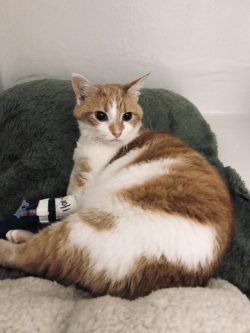
Sophie, an eight-year-old spayed female Domestic Short Hair cat presented to the Michigan State University Small Animal Clinic’s Orthopedic Surgery Service for evaluation of medial patellar luxation affecting both hind limbs, as diagnosed by her primary care veterinarian. Sophie’s owners first noticed that she was lame in her hind legs four months prior to presentation, when they found her unable to jump up onto the back of the couch and up onto their bed. They noted that occasionally she would bunny hop and appeared to be very stiff in the hind limbs when she got up from laying for a long time. They reported no issues with Sophie jumping down off of things.
On presentation, Sophie was bright, alert, and responsive. She tolerated her examination very well—and the same certainly cannot be said for all cats! She was appreciated to be moderately overweight but otherwise her general physical examination was within normal limits.
On orthopedic examination, no neck or spinal pain was appreciated. It was observed that the right patella could be completely luxated medially with digital pressure and remained temporarily luxated once the pressure was released. This was graded as Grade II/IV medial luxation using the traditional Putnam scheme or using the proposed feline grading scheme, as a grade B. The left patella was luxated medially without any manipulation but could still be replaced into the trochlear groove with digital pressure. This was graded as a grade III/IV medial luxation using the Putnam scheme or a grade D using the feline scheme. Positive cranial tibial thrust and cranial drawer were noted on the right stifle. The left stifle was stable in cranial drawer and cranial tibial thrust both conscious and under sedation. Both stifles were circumferentially thickened. The remainder of the examination was within normal limits.
Diagnosis
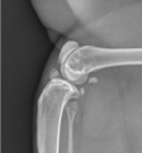
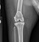
Bilateral stifle radiographs were performed. Opacification encroachment of the infrapatellar fat pad was evident bilaterally. The medial fabella was not mineralized on either side. A large mineralized fragment was evident cranially within the stifle joint on the mediolateral view of the right stifle; this finding has been associated with cranial cruciate ligament rupture in cats. A very small mineralized area was evident within the cranial stifle joint on the mediolateral view of the left stifle also. Mild periarticular osteophytosis was evident affecting the right stifle. The patella was appropriately situated on both craniocaudal views. At the periphery of the craniocaudal views the coxofemoral joint was evident and very mild remodeling of the cranial effective acetabular margin was evident bilaterally consistent with radiographically mild hip osteoarthritis. The stifle radiographs, in conjunction with the physical examination findings were consistent with bilateral degenerative joint disease affecting the stifle joints, with mineralization likely secondary to the degenerative process, although incidental mineralization could not be ruled out on the left. The degenerative disease was considered likely secondary to the medial patellar luxation and cranial cruciate ligament impairment on the right.
Sophie’s final diagnosis was bilateral medial patella luxation and concomitant right cranial cruciate ligament rupture. The recommended treatment was surgical stabilization of both conditions which Sophie’s owners were keen to pursue.
Treatment and Outcome
The combination of Sophie’s injuries is very uncommon in cats, and there is very little information about how to treat it. Sophie’s patellar luxations were considered very unlikely to resolve or improve substantially without surgical treatment. While some cats with cranial cruciate ligament rupture do improve clinically with non-surgical management, this improvement does take a long time and is often incomplete with stifle instability remaining. Therefore, surgery was considered to be the most efficient route to a full recovery. Exactly which surgery though, was the question.
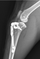
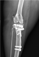
One potential option was use of an extracapsular stabilization technique for the cranial cruciate ligament component, combined with a tibial tuberosity transposition, block recession trochleoplasty, partial parasaggital patellectomy and soft tissue balancing for the patellar luxation. However, in dogs, an alternative technique, the tibial tuberosity transposition and advancement (TTTA) was recently shown to provide superior clinical outcomes with fewer associated complications. As such, the Orthopedic Surgery team decided to adapt the TTTA technique for use in cats. This is the first time that this has been reported to our knowledge.
Sophie was placed under general anesthesia. Exploration of the right stifle revealed a very shallow trochlear groove and a comparatively wide patella. A block recession trochleoplasty was performed to deepen the groove and a partial parasaggital patellectomy was performed to ensure that the patella could fit into the groove. The cranial cruciate ligament was partially torn, and the remnants were debrided. The medial meniscus was firm and torn and was partially removed. The lateral meniscus was within normal limits. A TTTA was performed using a 6mm cage, a 2-fork plate and a 4mm spacer. This is a surgery that changes the biomechanics of the knee (by creating a 90-degree angle between the tibial plateau and the straight patellar ligament) so that the function of the cranial cruciate ligament is no longer necessary for stifle stability. It also treats the patellar luxation by straightening the quadriceps mechanism. Minor adjustments to the technique and implants used in dogs were necessary to facilitate application in a cat but the procedure went very well.
Postoperative radiographs revealed satisfactory implant positioning and patellar tendon angle. Sophie recovered well from anesthesia. She remained at the Hospital overnight and was up and about, putting weight on the limb less than 24-hours postoperatively.
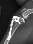
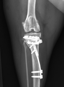
On discharge, Sophie’s owners received instructions for incision care, activity limitations, physical rehabilitation, diet alterations, and recheck appointments. The prognosis for a return to full function was considered very good, but strict exercise restriction would be imperative in avoiding complications such as implant breakage and since the day she returned home from the Hospital. The only abnormalities noted were a very mild and intermittent limp occasionally and that she had a tendency to sit with her right hind limb extended rather than flexing it underneath her. The night prior to presentation she had been jumping between two beds after escaping from her crate but no adverse effects from this had been noted. Her wound had healed well; no complications were noted, and she was no longer receiving any analgesic medications.
A follow-up orthopedic examination revealed an occasional mild weight-bearing lameness on the right side, but this was not evident with every step. At stance, she was weight-bearing evenly and she was able to jump up on to low surfaces in the consulting room with no difficulty, in fact she jumped up on to a chair to look out of the window when visiting the MSU Small Animal Rehabilitation Clinic. Orthopedic examination revealed no pain upon palpation over the implants (although these were easily palpable under the skin). The right stifle was stable in cranial tibial thrust and the patella could not be luxated. As detailed previously, the left patella was luxated medially, but the grade of luxation was unchanged from initial examination and the stifle remained stable in cranial tibial thrust.
Radiographs of the right stifle revealed that healing of the osteotomy site was ongoing with organization of the previously placed graft and bridging obtained at some areas but not along the entire length of the osteotomy. This was considered satisfactory progress for this stage postoperatively. There were no signs of implant-associated complications.
At this stage, it was recommended that Sophie return to a relatively normal activity level. However, boisterous play with other cats or jumping on to very high surfaces continued to be restricted. Once the bone has completely healed, she will be released from these restrictions.
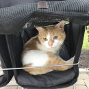
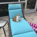
At present, six months post-operation, Sophie is reported to be doing very well. She traveled south to Florida with her owners, where she enjoys long walks in her stroller and sunbathing.

