By Karen Perry, BVM&S, MRCVS, DECVS
History and Diagnosis
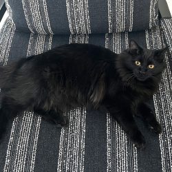
In 2014, Abbey, an 11-year-old spayed female Domestic Short Hair, began displaying signs of lameness, especially while walking or jumping. Her owner was unaware of any traumatic event that would have caused an injury. In 2015, Abbey’s primary care veterinarian prescribed subcutaneous Adequan injections every four-to-eight weeks and Glycoflex II chewable tablets every day. One year later and after little progress, Abbey was referred to the MSU Veterinary Medical Center’s Rehabilitation Service for a gait abnormality evaluation.
Dr. Sarah Shull found Abbey to have severe weight bearing lameness on both thoracic limbs and a short, choppy, stilted gait on both pelvic limbs. Dr. Shull also noted left and right carpal hyperextension. Abbey had moderate-to-severe muscle atrophy over the scapula and humerus of both thoracic limbs. Elbow flexion caused discomfort, as did extension on both thoracic limbs.
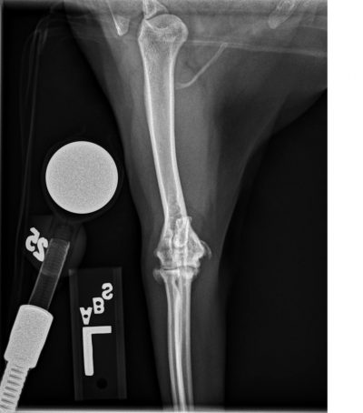
While the physical examination indicated osteoarthritis, Dr. Shull referred Abbey to Dr. Karen Perry of the Orthopedic Surgery Service for further diagnostics and radiographs. The physical examination, radiographs, and a CT scan revealed a second and third diagnosis: medial humeral epicondylitis (MHE) and osteochondromatosis.
Treatment and Outcome
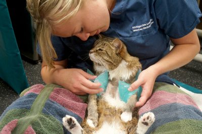
After four weeks of medical management using Robenacoxib pulse therapy—three days on, one day off—to combat inflammation, only very mild improvement was noted and surgery was deemed to be necessary to improve Abbey’s quality of life. Dr. Perry made an approach to the medial humeral epicondyle on the left to remove the new bone associated with the MHE, which was within the humeral head of the flexor carpi ulnaris muscle. This bone was found to be compressing the ulnar nerve, which was adhered to surrounding structures secondary to chronic inflammation, and this likely explains at least part of the reason why Abbey had failed to respond to medical management. An elbow arthrotomy also was performed to remove the three large mineralized intra-articular bodies that had been noted on CT. Severe cartilage damage (Outerbridge grade three) was evident affecting the medial coronoid process in the left elbow. Abbey’s right elbow was less severely affected, but two small mineralized bodies were removed from the joint and Outerbridge-grade-two cartilage damage was noted. A carpal flexion bandage was placed on the left to protect the repair of the flexor carpi ulnaris muscle for the first two weeks after surgery.
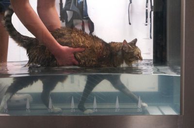
Immediately following surgery while Abbey was still under anesthesia, Dr. Perry worked with the Rehabilitation team to apply laser therapy, icing, passive range of motion exercises, and acupuncture. Bi-weekly visits with the Rehabilitation team were scheduled for the next six weeks to continue these therapies. Abbey progressed well over the next three weeks and had positive rechecks with the Orthopedics team, so Dr. Shull added massage and hydrotherapy to Abbey’s regimen. For her first session, Abbey walked on the underwater treadmill through seven inches of water for one and two-minute bursts with rest periods in between. Today, Abbey continues to improve and progress at each therapy session. Since surgery, it has been possible to wean Abbey off of all medications and she is significantly more active. Abbey will continue postoperative rechecks with the Orthopedic team. While Abbey’s osteoarthritis will progress over time, removing the mineralizations likely will help reduce the speed at which it progresses, as well as resolve the compression on the ulnar nerve, both of which will improve her comfort level and quality of life.
Comments
Medial humeral epicondylitis is caused by avulsion and calcification of the flexor tendons at their insertion site on the medial humeral epicondyle. Cats with MHE can present with or without concomitant arthritis and other changes, including osteochondromatosis, as in Abbey’s case. Medial humeral epicondylitis has been associated with subluxation of the humeroulnar and humeroradial joints, and by extension, this may cause incongruity and subsequent cartilage defects. The bony spurs and free fragments associated with MHE also can cause separation of the humeral and ulnar heads of the flexor carpi ulnaris muscle and impinge on the ulnar nerve, which sits in a confined space between these muscles, causing discomfort that can be difficult to manage medically. The cause of MHE is essentially unknown, but may be related to trauma or repetitive overuse causing partial or complete avulsion of the flexor muscles leading to tendinosis.
Cats may be prone to MHE due to their active lifestyles, and often manifest a pain response on digital palpation over the flexor muscles and medial epicondyle. Carpal flexion also elicits pain in the majority of these cases. Both of these findings were present in Abbey.
In the earliest stages of MHE, inflammation only is present with fibrosis and calcification becoming evident at the later stages. Therefore, MHE may be overlooked in the early stages, as there will be no signs on radiographs. In mild, chronic cases, radiographic signs are subtle with rounding and slight irregularity of the medial humeral condyle, which was seen in Abbey’s right elbow. In moderate chronic cases, pronounced irregular new bone formation with bony spur formation is seen over the medial humeral condyle. In advanced, severe cases, large semicircular mineralized structures may be seen at the caudomedial aspect of the elbow joint, which is what was seen on Abbey’s left elbow.
Mild cases of MHE can be treated conservatively using environmental modulation, physical therapy, weight reduction, and analgesia. In cases where conservative management is ineffective, treatment consists of excision of the pathologic portion of the tendon and repair of the resulting defect. Ulnar nerve adhesions or compression also must be recognized and treated appropriately. Any intra-articular mineralizations can be removed during the same procedure. A carpal flexion bandage is recommended to reduce tension on the repair postoperatively.
Most cats treated surgically for MHE show significant improvement in lameness and pain level post-operatively, and the short-term outcome is fair to good. Recurrence of a bone spur or mineralization may occur, but the likelihood of recurrence is not known. Information and literature about the long-term outcome following surgical treatment of MHE is currently not available as cats have not been followed for a long enough time period post-operatively to assess the true rate of recurrence.
For more information about MHE in cats, please reference Dr. Perry’s paper “The Lame Cat: Common Culprits of Non-Traumatic Lameness when Pain Localises to the Elbow or Shoulder” published in volume 20 (2), 2015, of Companion Animal, pages 86–91.

