Featuring Maureen Spinner, Lauren Meneghetti, Sara Jablonski, Paige Albers, Annette Petersen, Erica Noland
This story features Dr. Maureen Spinner, Dr. Kelly Schrock, and Dr. Lauren Meneghetti, Soft Tissue Surgery; Dr. Sara Jablonski and Dr. Paige Albers, Internal Medicine; Dr. Annette Petersen and Dr. Erica Noland, Dermatology
History and Presentation
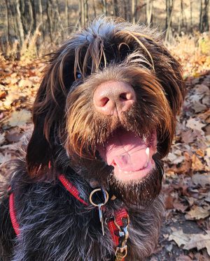
Otto, a one-year-old Wirehaired Pointing Griffon, presented to the Internal Medicine service at the MSU Veterinary Medical Center after his primary veterinarian noted that the energetic young dog did not appear to have an opening for his right ear to the ear canal.
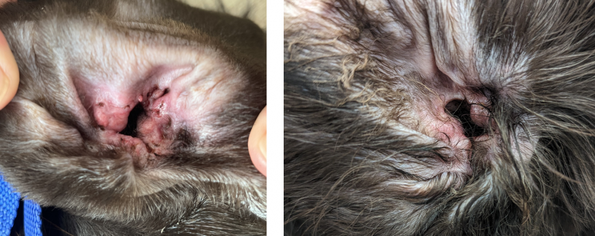
Otto demonstrated no clinical signs associated with pain, irritation, or pruritus of the right ear. Physical examinations at MSU conducted by the Internal Medicine and Dermatology services suggested that Otto had a birth defect resulting in incomplete formation of the external opening of the ear canal.
A CT scan confirmed that the vertical and horizontal components of the ear canal were present, but there was no connection to the outside of the ear. Further, the right vertical and horizontal ear canal, as well as the bulla, were filled with what was suspected to be normal hair, skin, and oils produced by the ear epithelial lining.
With no normal ear canal opening, surgery was recommended to address Otto’s ear abnormalities and further collection of the material within the ear canal. Without surgery, the buildup of material within the ear would likely damage the middle and inner ear structures, as well as increase Otto’s risk of infection, pain, and potential neurologic changes.
Surgery and Treatment
A few surgical options were discussed:
- Resection and anastomosis of the ear canal to create a complete and functional right ear.
- Lateral ear canal resection to remove the vertical portion of the ear canal, creating a new opening on the side of Otto’s head, just below the base of the ear pinna. This procedure is accompanied by increased risk for future ear infections.
- A total ear canal ablation and bulla osteotomy (TECABO) to remove the ear canal entirely. Because of complications associated with this procedure, it was not strongly recommended for an otherwise healthy young dog.
Otto’s family elected an ear canal resection and anastomosis procedure to give Otto the best possible chance at retaining full use of his ear. The surgery was completed by MSU’s Soft Tissue Surgery service.
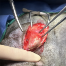
1. An incision was made over the blind ended right ear canal, which was located caudal to the pinna and both visible and palpable through the skin. A combination of blunt and sharp dissection with Metzenbaum scissors along with monopolar electrocautery were used to partially dissect out the dorsal portion of the vertical canal by carefully trimming away the subcutaneous tissue and muscle. A Gelpi retractor was used to aid in exposure during dissection and identification.
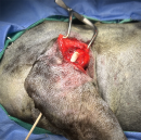
2. A #15 scalpel blade was used to create a perforation for the future external ear canal opening. A full thickness incision was made, and associated skin and cartilage was removed to create the new stoma for the ear canal. A sterile cotton tip applicator can be seen passing from the newly created stoma towards the site of the exposed vertical ear canal.
3. A new #15 scalpel blade was used to sharply incise the blind end of the vertical ear canal to visualize within the canal. A combination of suction and mosquito hemostats were used to remove hair and ceruminous material from the canal. The canal was then lavaged with sterile saline and actively suctioned.
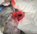
4. The now-open vertical canal was advanced under the subcutaneous tissue to exit the created pinnal opening with the use of 4-0 nylon stay sutures.
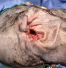
5. Anastomosis was then performed using 4-0 Monocryl interrupted sutures to achieve good apposition. The skin incision over the vertical ear canal was routinely closed.
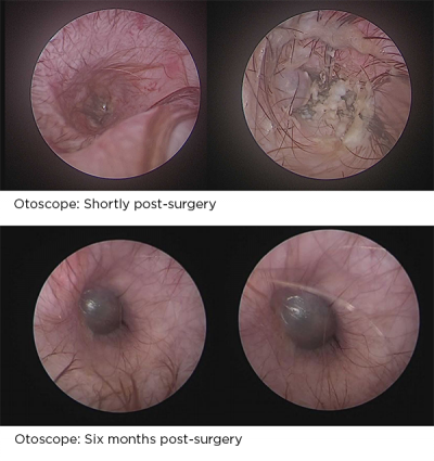
Following surgery, Otto underwent otoscopic evaluation by the MSU dermatology service to assess the ear canal integrity. Additional hair and debris were removed and the ear drum (tympanic membrane) was identified to be intact, but flaccid in appearance. Ear canal swabs were collected for microscopic evaluation and Otto recovered well from surgery.
Recovery
Otto’s incisions healed well. He was later treated by the MSU Dermatology service for yeast infection of both ears. Otto was treated with a topical anti-fungal and the infection resolved routinely.
Otto was re-evaluated approximately 5 months following surgery and was found to be doing well at home. His owner reports that his ability to hear seems as normal as any other dog.
