A novel surgical operative system and method have earned Michigan State University another US patent. Dr. Loïc Déjardin’s latest invention will make surgeries for injured sacroiliac joints—which is where the spine connects to the pelvis—easier, faster, and safer.
Surgery with SILIS-MILAD

Déjardin, professor and head of Small Animal Orthopedic Surgery at the MSU Veterinary Medical Center, invented the Sacro-Iliac Luxation Instrumentation System (SILIS) and a Minimally Invasive Lucent Aiming Device (MILAD). When compared to current methods, the SILIS-MILAD better and more quickly provides accurate and reliable reduction and stabilization of a dislocated or fractured sacroiliac joint. The SILIS-MILAD also allows surgical teams to virtually eliminate their exposure to radiation and dramatically reduces it for patients.
“The SILIS-MILAD is an innovative, effective, and reliable operative system designed to enable surgeons to improve minimally invasive reduction and fixation of sacroiliac joint luxation and fracture. This system prevents implant malposition during fixation and improves safety for operating personnel and patients during surgery. With the SILIS-MILAD, we are reducing surgical morbidity and post-operative complication rates, and thus, we are improving clinical outcomes,” says Déjardin, who also is the Wade O. Brinker Endowed Chair of Veterinary Surgery at MSU and an ACVS founding fellow in Minimally Invasive Orthopedic Surgery.
Surgical History and a New Way Forward
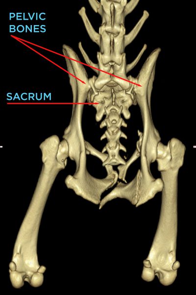
Sacroiliac joint luxations, or dislocations, and fractures (SIL/F) are often seen in dogs and cats who have suffered road traffic accidents. Traditionally, a surgeon would use extensive gluteal dissection and temporarily move the ilium bone to access the sacrum bone. The surgeon would drill a hole through the sacrum, return the ilium to its original position, and use a screw to secure the ilium to the sacrum.
This surgical approach, called open reduction and internal fixation (ORIF), has an inconsistent success rate, in part due to the invasive surgical approach and complications that result from suboptimal screw placement.

“The complexity of the ORIF procedure and risk of complication often deter veterinarians from choosing this surgical option. Instead, these dogs are too often ‘cage rested’ to heal, but this unnecessarily prolongs their pain and recovery,” says Déjardin.
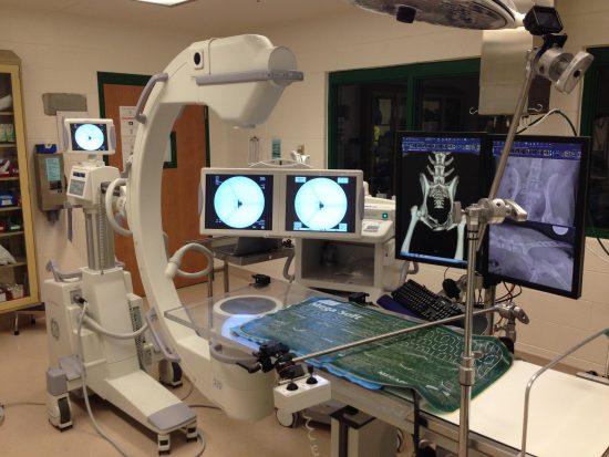
Eventually, a minimally invasive osteosynthesis (MIO) procedure was developed by Professor James Tomlison to improve screw placement accuracy and reduce complications. With MIO, an intraoperative imaging modality called fluoroscopy creates a real-time “X-ray movie” of the surgical site. The surgical team then uses these X-rays to reduce the joint from the outside, without direct access to the ilium. Then, a temporary pin holds the ilium in place while the surgeon inserts an iliosacral screw to permanently secure the joint. While this approach offers more reliability than ORIF, maintaining reduction and providing accurate screw orientation remains a challenge. In addition, MIO exposes both the patient and surgical team to ionizing radiation.
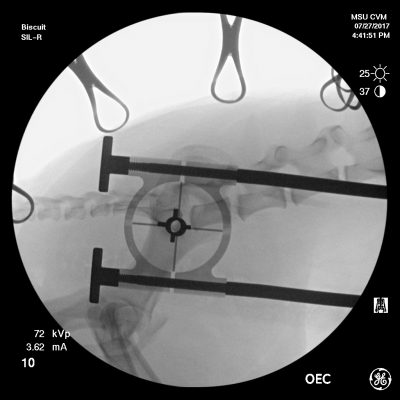
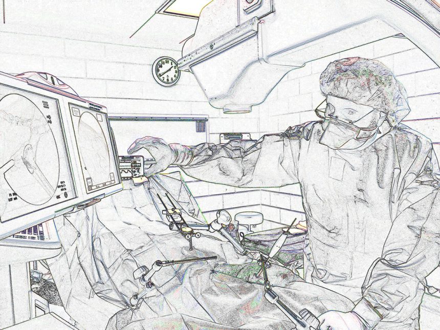
The SILIS-MILAD solves these problems. It features moveable and lockable articulated arms—one or two for reduction and one for fixation—that latch onto a surgical table. The free end of each arm is coupled to either a handle, used to manipulate bones (reduction arm), or the MILAD, a radiolucent drill guide used to provide accurate screw placement in the sacrum (fixation arm).
To minimize surgical trauma, Déjardin uses small incisions to insert the reduction handle(s) to the ilium. Following reduction, the arm is locked in place to immobilize the ilium. Next, Déjardin positions the MILAD above the surgical site. He inserts it through the gluteal muscles via an additional small incision, oriented along the sacral axis, and finally, locks the fixation arm in place.
“Because the sacrum is a very small bone, it is critical that the iliosacral screw be placed accurately along the sacrum’s safe implantation corridor, for which there is almost no room for error,” says Déjardin.

Throughout the procedure, Déjardin and his team step away from the fluoroscopic unit while radiographs are taken. Déjardin reviews the radiographs to check reduction until alignment has been restored and optimal screw placement has been achieved for fixation. Through this process, and unlike conventional MIO of SIL/F, the SILIS-MILAD virtually eliminates radiation exposure to the surgical and anesthesia team while providing stable reduction and accurate fixation.
While today’s typical SIL/F surgery takes between one and two hours, Déjardin routinely spends 20 minutes or less on each of his patients.
“The SILIS-MILAD increases surgical speed, accuracy, and consistency,” says Déjardin. “And not only are we able to protect OR personnel from exposure to radiation, but we also reduce exposure for the patient.”
Déjardin says the SILIS-MILAD may have other surgical applications, too. He and his collaborators see the potential for this instrumentation to assist in other procedures like long bone fractures and total joint replacements as robotic-assisted surgery continues to evolve.
“It’s easy to imagine how the SILIS-MILAD arms could be replaced by robots to offer improved accuracy, reliability, patient outcomes, and cost effectiveness.” In the meantime, Déjardin believes that the SILIS-MILAD technology could be applied across species. “Because veterinary and human medicine are inextricably connected, we have high hopes that the positive impact the SILIS-MILAD’s made on our animal patients will one day be expanded to people.”
To learn more about the SILIS-MILAD and veterinary orthopedic research at the MSU College of Veterinary Medicine, contact the Déjardin Laboratory.
About the Inventor
Loïc Déjardin, DVM, MS, DACVS, DECVS, is the Wade O. Brinker Endowed Chair of Veterinary Surgery. He is a professor and head of Small Animal Orthopedic Surgery at Michigan State University and an ACVS Minimally Invasive Small Animal Orthopedic Surgery founding fellow. A graduate from Toulouse – France, Déjardin then completed his Surgical Residency, then MS, with Dr. Steven Arnoczky at MSU.

Déjardin has authored approximately 90 research proposals (which total approximately $7 million), created 8 inventions, and holds 3 patents on orthopedic implants and surgical devices. He has received several prestigious awards in both veterinary and human medicine, as well as in engineering including the O’Donoghue Sports Injury Research Award (AOSSM), the Zandman Award (Soc. Exp. Mechanics), Distinguished Postdoctoral Veterinary Alumnus Award (MSU), and the Pfizer-Zoetis Award for Excellence in Research. His publications include approximately 160 peer-reviewed scientific papers and abstracts, 20 book chapters, and approximately 475 presentations in the US, Europe, Latin America, and Asia.
As an AO Foundation International Faculty and former Trustee committed to continuing education worldwide, Déjardin regularly speaks at national and international meetings and courses. He started a Minimally Invasive Osteosynthesis program at MSU in the early 2000s and developed a novel interlocking nail suited for MIO, as well as a new technology devised for the MIO of sacroiliac luxations. Since 2009, Déjardin has created and chaired the first comprehensive AOVET Master Course on MIO.
His clinical interests include comparative orthopedics, traumatology, MIO, and revision surgery, as well as total joint replacement. His current research activity focuses on biomechanics, implant and instrument design, and total joint replacement (elbow, hip, knee, ankle), as well as robotics and kinetics.
- Read more about Dr. Loïc Déjardin.
- Déjardin’s publications about SILIS-MILAD (“Apparatus and Method for Minimally Invasive Osteosynthesis of Sacroiliac Luxations/Fractures,” US patent 10,687,863 B2, issued June 23, 2020):
- Dejardin et al., “Minimally invasive lag screw fixation of sacroiliac luxation/fracture using a dedicated novel instrument system: Apparatus and technique description,” Veterinary Surgery,” 2017: 1-11 (2017).
- Dejardin et al., “Comparison of open reduction versus minimally invasive surgical approaches on screw position in canine sacroiliac lag-screw fixation,” Vet. Comp. Orthop. Traumatol., 29:290-7 (2016).
- Past stories about the SILIS-MILAD:
The Impact of Mentorship
In addition to his research and clinical work, Dr. Loïc Déjardin is an active mentor to the next generation of orthopedic surgeons. “Loyalty and dedication are essential to successful mentorship. Early recruitment of young talent and continuous nurturing and encouragement of your mentees are key to any mentoring relationship. It is important to recognize that this is a lifelong endeavor. While this commitment takes time and energy, the success of your mentees is most rewarding” says Déjardin.
Three of Déjardin’s mentees are co-authors on his SILIS-MILAD research:
- Laurent Guiot, DVM, DACVS, DECVS, joined Déjardin and the MSU College of Veterinary Medicine in 2006 to pursue an international surgical fellowship, a specialization program initiated by Déjardin in the early '90s. Following his three-year residency with Déjardin, which focused on orthopedic surgery, Guiot became an assistant professor of small animal orthopedic surgery at MSU. Guiot became a boarded surgeon of the American and European Colleges of Veterinary Surgeons in 2011.
- Reunan Guillou, DVM, DACVS, joined the MSU College of Veterinary Medicine in 2007 as a research associate in Déjardin’s laboratory. Guillou furthered his clinical training as one of the College’s surgical fellows, and then completed a surgical residency, with an emphasis on orthopedic surgery with Déjardin. After achieving board certification in 2013, Guillou joined MSU’s faculty as an assistant professor.
In 2013, Guiot and Guillou were recruited to lead the creation of a new orthopedic surgery facility for The Ohio State University in Dublin, Ohio. Eventually, they moved to Culver City to co-develop an orthopedic service for a chain of California-based animal hospitals.
- Danielle Marturello, DVM, MS, DACVS, is an assistant professor of orthopedic surgery at MSU. Marturello completed a research associateship in Déjardin’s laboratory in 2014 and then her surgical residency training, with a dual Master of Science degree, under Déjardin. Her work focused on the development of a feline bone substitute and mechanical testing of feline orthopedic implants, which has been presented at both national and international meetings. Dr. Danielle Marturello, Dr. Karen Perry, and Dr. Loïc Déjardin also were the first to publish clinical results from the feline I-Loc, an interlocking nail implant developed by Déjardin. Dr. Marturello is particularly interested in traumatology and biomechanical testing of orthopedic implants. Her current research activities are focused on 3D printing, with interest in the development of patient-specific instrumentation and implants.
For their outstanding and impactful work in applied basic science and clinical research, Marturello, Guillou, and Guiot received numerous national and international accolades from the American and European Colleges of Veterinary Surgeons, as well as the Veterinary Orthopedic Society. Such praises include the prestigious Mark S. Bloomberg Memorial Resident Research Award.
By E. LaClear
