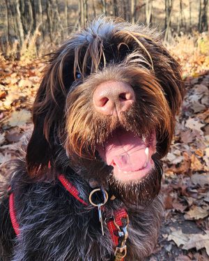
Otto, a one-year-old Wirehaired Pointing Griffon, seemed like any other energetic young dog. He played, he barked, he responded when called—no problem.

But one long holiday weekend, his owners noticed unusual ear-related symptoms. “He had the typical signs of an ear infection: scratching at it, shaking his head a lot,” his owner said.
The family took Otto to his primary veterinarian, who confirmed their suspicions. But then came a surprise.
“She does the exam and says to us, ‘Yup, he’s got an ear infection in his left ear. But has anyone ever talked to you about his right ear?’”
Otto's owners were confused.
“Our veterinarian explained that she didn’t think Otto had an ear canal in his right ear, and that she’s never seen or heard of that before,” they explain.
Even though Otto had shown some ear discomfort over the weekend, he had never exhibited signs of muffled hearing or discomfort—anything that would indicate he was missing a crucial part of the body.
“He was a young dog, so sometimes not listening or knowing when to follow commands just seemed like part of raising a puppy,” says his owner. “He was always (and continues to be today) better with hand signals than verbal commands.”
The veterinarian referred Otto to the Internal Medicine service at the MSU Veterinary Medical Center for a more detailed examination.
Hear We Go: Building an Ear
Veterinary professionals within MSU’s Internal Medicine and Dermatology services took a deep look at Otto’s ear, using physical exams, a video otoscope tool, and a CT machine to confirm: Otto had a birth defect that meant the external opening of the ear canal never fully formed. This condition, known as ear canal atresia, is rare.
His MSU medical team explained that while Otto could probably hear in the impacted ear, sounds were muffled, akin to being underwater. His other ear would have functioned normally. And though a missing ear canal did little to impact Otto’s hearing and overall wellbeing, the abnormality led to a buildup of debris within the young dog’s head, which in time would potentially damage his middle and inner ear structures, as well as risk infection. Surgery would be needed to prevent future complications.
Spartan surgeons discussed possible procedures with Otto’s family, ultimately deciding on the option that promised the most positive long-term impact on Otto’s health: a resection and anastomosis of the ear canal. In other words, to create an opening from scratch.

The surgery was completed by MSU’s Soft Tissue Surgery service. While Otto was monitored by members of the Anesthesia service, surgeons carefully made an incision and removed skin and cartilage from the area, creating what is known as a stoma, or a surgically-made hole. The stoma connected Otto’s previously blocked ear canal to the outside of his body. From there, the medical team cleared out the material that had been trapped within. A process called anastomosis was conducted to position the new opening properly alongside the ear canal, and all incisions were sewn neatly.
Otto recovered well from anesthesia, and his surgical site began to heal into a clean new ear opening.
Recovery
About five months following surgery, Otto returned to MSU to report that he was doing well at home.
“After the surgery, I do think he hears a lot better!” says his owner. “He became more engaged with me when I said his name or gave him a command. I didn’t have to try as hard to get his attention. Of course, that could also be related to him becoming more mature, so it’s hard to say for certain.”
Though his hearing now seems normal, Otto prefers to use a different sense. “Otto is a hunting breed—and his favorite thing to do is use his nose!” says his owner. “We’ve become very involved in scentwork, a dog sport where we work together to find a hidden odor. It’s so much fun for both of us!"


