Simulation Models at the College
- Dog and cat jugular and leg venipuncture and IV catheter placement
- Horse venipuncture and neck intramuscular injection
- Intramuscular and subcutaneous injection practice pads
- Surgical instrument handling task trainers
- Cow tail for blood draw
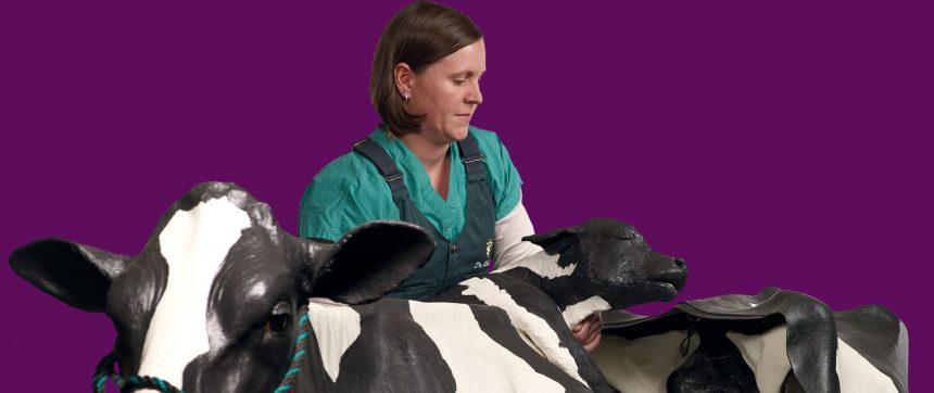
- “Mootilda” cow dystocia trainer with two calves
- Equine colic simulator
- Realistic skin practice pads for suturing
- Urinary bladder for cystocentesis (sampling urine through needle)
- Female dog urinary catheter placement model
- Dog endotracheal tube placement model
- Lymph node aspirate
- Cat castration model
- Duo chest tap and pericardial tap model
- Stuffed animal patient used for surgical draping practice
In medical education, hands-on experience is crucial for students to go on and become successful veterinarians. But before working with real patients, simulation models provide a safe, risk-free environment in which students can practice.
The MSU College of Veterinary Medicine prioritizes high-quality education by providing simulation models to students. The College recently added to their collection by obtaining three life-size simulation models—two equine and one bovine. Professors use these realistic educational tools to provide students with hands-on experience with clinical situations in a low-stakes, low-pressure environment. Before working with live animals, DVM and veterinary technology students practice procedures and treatments, which may include sutures and different types of physical examinations.
“Simulation models are key for our students to develop fine motor skills and basic techniques,” said Dr. Jen Roberts, assistant professor in Large Animal Clinical Sciences. “This practice makes real-life situations and critical thinking more natural and automatic.”
The new equine colic simulator is modeled after a 15-hand quarter horse and will be a tool in teaching the various complications of colic, why they occur, and how they can be solved. The equine theriogenology model will be used to simulate physical examinations of the reproductive system. The bovine theriogenology model includes five different interchangeable reproductive tracts—two non-pregnant and three pregnant—to teach students about the different stages of pregnancy.
In addition to providing practice in clinical scenarios, the models increase teaching efficiency as students work independently to develop skills with repeated practice.
“A clinical skills lab offers the students a chance to walk in and practice at convenient times for them,” said Bea Biddinger, licensed veterinary technician, who oversees open hours in the junior surgery lab for clinical skills practice.
The College is known for graduating practice-ready students and is working to provide even more clinical experience to students. Currently, second- and third-year students practice on the simulation models. Dr. Roberts is working with the College’s Curriculum Reinvention team to put students’ hands on the models earlier in their preclinical veterinary education.
“The sooner we can get our students to learn and practice these simulations, the more prepared they will be,” said Dr. Roberts.