History and Presentation
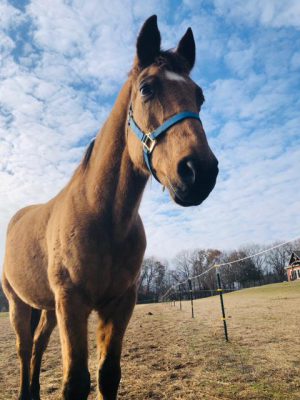
Remington, a 16-year-old Quarter Horse gelding, presented to the Michigan State University Large Animal Clinic for further evaluation of severe colic signs that were non-responsive to management on the farm where he lives. The patient was initially noted to be acting colicky at noon that day, at which time he was laying down while out in the field. Remington’s clinical signs progressed to rolling and attempting to lay down while being walked.
A dose of an oral anti-inflammatory (Banamine) was administered at 12:30 p.m. that day. Remington’s primary care veterinarian was called out to evaluate him on site. On evaluation, he had a heart rate of 70 and was breathing heavily and sweating. On rectal examination, the veterinarian could not get past the pelvic canal and noted to have felt something like a blind-ended pouch. Remington required heavy sedation for evaluation. Given the findings, the primary care veterinarian recommended referral to the MSU Large Animal Clinic for further assessment and treatment. Butorphanol was administered for the trailer ride.
Diagnosis
On presentation, the MSU veterinary healthcare team performed a full evaluation, including physical examination, ultrasound, rectal examination, and nasograstric intubation. The ultrasound revealed an empty stomach with margins at approximately 11–12th intercostal space. Both of Remington’s kidneys were visualized, though the left was difficult to identify due to the presence of gas within the intestine in that area. The spleen was mildly displaced ventrally in the left lateral abdomen but didn’t extend into the right abdominal quadrants. No distended loops of small intestine were seen, and the large colon appeared of normal wall thickness.
No net reflux was present when the NG tube was placed. However, there was a moderate amount of dry feed material present within the stomach. The stomach was lavaged with 16 L of water.
Upon rectal examination, the rectal walls were contracted down to a finger-size opening with a fecal ball palpated on the other side of the stricture at about 10–12 inches in from the rectum. Cranial and ventral to the stricture, a 4–6 cm diameter round structure was palpated. This was suspected to be a strangulating lipoma, which caused the stricture. The stalk of the lipoma was easily palpated but too thick to dislodge digitally through the small colon.
Based on these findings, the MSU Equine Emergency Service diagnosed Remington with a strangulating lipoma of his small colon.
Treatment and Outcome
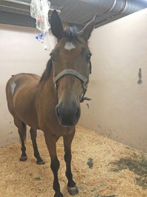
An IV catheter was placed in Remington’s left jugular vein and he was sedated for another rectal examination. Initially, an abdominal exploration under general anesthesia was recommended to remove the lipoma and release the strangulated small colon. During the preparation for surgery, Remington’s owner called the MSU Large Animal Clinic to reconsider surgery for the patient. The owner had decided to rather proceed with humane euthanasia.
At that time, given the known location of the lipoma, a localized approach via standing sedation and local anesthesia was offered as a possible option. Remington’s owner was informed that the standing procedure would only allow getting to the lipoma to remove it and that a full abdominal explore could not be performed through this approach. If Remington had additional diseased areas of the GI tract, these could potentially be missed. Also, intra-operative and post-operative complications like hemorrhage, damage to the GI tract, surgical site infection, peritonitis and others were discussed.
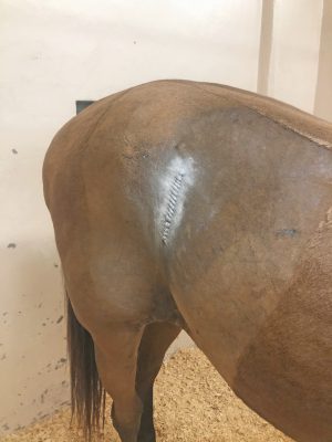
Remington’s owner consented to the procedure, knowing the intra-operative and post-operative risks. A right flank laparotomy was performed through a line block with a local anesthetic (Carbocaine). The lipoma was identified in the caudodorsal quadrant. The stalk was identified and transected with scissors guarded by the surgeon’s hand and the lipoma was removed from the abdomen. A doughy impaction of the small colon oral to the strangulation was palpated during surgery. The large colon also had some doughy feed material. No other abnormalities were identified. The abdomen was checked for major hemorrhage after resection of the lipoma stalk. Once no major bleeding was identified, the incision was closed in three layers. After the procedure, a rectal examination was performed to confirm the patency of the rectum/aboral small colon.
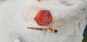
The procedure went without complication and Remington was continued on medical therapy post-operatively. Remington developed mild post-operative ileus between 12- and 24-hours post-op but improved greatly by 36 hours post-op. He was started on a re-feeding program after that and was slowly weaned off all medications and supportive care. He had been doing well in the Hospital since and was sent home on continuous monitoring. He was sent home with specific dismissal information, including instructions for how his owner should monitor, feed, care for his incision, and house and exercise him.
Five months post-operatively, Remington’s owner has reported that he’s doing well. He noted that since surgery, Remington is better mannered and more friendly. He no longer lays down as much and stands better for the fairer.
Comments
A lipoma is a fatty tumor most commonly located on the mesentery of the GI tract in horses. While in dogs, it is most often situated between the skin and the underlying muscle layers. It is slow-growing and is not malignant, nor is it normally harmful. In horses, as the tumor grows from its stalk, its increasing weight causes it to hang and has the potential to loop around a section of the intestine. A lipoma doesn’t always result in an immediate problem unless the loop tightens. If this occurs, it prevents ingested material from passing and cuts off the blood supply to the compressed tissue. The horse then begins to show signs of colic, and discomfort may become severe.
The most common treatment for lipomas in horses is surgical removal on an emergency basis, as they will not go away on their own or with medical treatment for colic. Recurrences after removal are uncommon.
A standing, localized approach was recommended in this case as a last resort option given that the exact location of the lipoma was known, and the patient was standing quietly while sedated and not wanting to go down and roll which made him amenable to standing surgery. A majority of horses with strangulating lipomas of their small intestine have unrelenting pain and will want to lay down and/or roll even with heavy sedation on board. Standing flank laparotomy is not a typical procedure performed on this type of case due to the high risk of complications that can stem from it but also because in the majority of cases with strangulating lipomas, a full abdominal explore is warranted to offer a better-guided prognosis post-operatively. We are thankful that Remington’s owner trusted us to give his horse a last-resort chance through this procedure and that despite minor complications post-operatively the patient ultimately did well and was able to go home to his family.

