History and Presentation
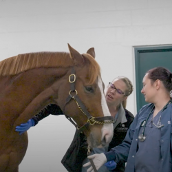
Rouby, an 18-year-old Oldenburg multiparous broodmare, was presented to Michigan State University’s Equine Emergency Service for dystocia that was unable to be resolved on the farm. She was two weeks overdue with her foal. Labor started earlier that day, but there was little progress. Rouby’s primary care veterinarian and their associate both tried to deliver the foal on the farm, but unfortunately, they were not able to make any progress.
On presentation, Rouby was bright, alert, and responsive. Vital parameters (temperature, heart rate, and respiratory rate) revealed tachycardia and tachypnea, likely secondary to discomfort. Mucus membranes were pink and tacky, with a capillary refill time of two seconds. Cardiothoracic and gastrointestinal auscultation were within normal limits. Digital pulses were clinically unremarkable. Fetal membranes were noted protruding from her vulva.
Diagnosis
Abdominal ultrasonography revealed no evidence of a fetal heartbeat or free peritoneal fluid. A sedated vaginal examination noted that the fetus was non-responsive to stimuli and was determined to be in anterior, longitudinal, dorsosacral presentation with the fetal head and neck retroflexed to the left with a 180-degree rotation causing the mandible to be dorsal to the maxilla.
Hemorrhage was noted during the vaginal examination, but an obvious uterine or vaginal laceration was not identified. Baseline hematology revealed evidence of mild dehydration and mild ionized hypocalcemia with no other clinical abnormalities.
Treatment and Outcome
Based on the fetal presentation with no evidence of a live fetus, controlled vaginal delivery under general anesthesia was recommended and elected. An intravenous catheter was placed, and the mare was administered broad spectrum antimicrobials and an anti-inflammatory pain medication.
Rouby was induced under general anesthesia and placed in Trendelenburg position with her hind limbs suspended from an overhead hoist. Numerous interventions were performed to alter fetal positioning. Following repulsion, mutation, and rotation, the fetus was eventually delivered vaginally. Initially, the colt did not have a corneal reflex, but a heartbeat was noted while ligating the umbilicus. The heart rate was low, so epinephrine was administered to increase the rate and force of cardiac contraction and improve tissue perfusion. The foal was intubated and provided supplemental oxygen while an intravenous catheter was placed in the left jugular vein.

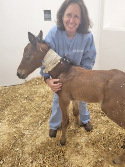
After 10–15 minutes, the foal self-extubated and was attempting to stand. A red rubber catheter was placed in the right nostril for nasal insufflation, and a nasogastric feeding tube was placed in the left nostril for colostrum administration. The foal was placed in a stall while Rouby recovered from general anesthesia, and he was administered a broad-spectrum antimicrobial intravenously and colostrum via the feeding tube. Rouby recovered from general anesthesia uneventfully and was introduced to her colt, who was able to stand without assistance at that time. A small amount of amnion was placed on the colt’s back to facilitate the bonding of mare and foal. Rouby accepted the foal and allowed him to nurse readily. The colt was closely monitored to ensure proper hydration and was maintained on intravenous antimicrobials due to the prolonged delivery and increased risk of sepsis.
Overnight, Rouby became progressively more tachycardic, prompting re-evaluation. Repeat hematology revealed moderate dehydration with a packed cell volume of 55% and a total solid of 7.8 g/dL, despite being 42% and 6.9 g/dL just 2 hours earlier following recovery from general anesthesia. Repeat abdominal ultrasonography was clinically unremarkable at that time with no evidence of increased free peritoneal fluid. Rouby was administered a bolus of intravenous balanced crystalloid fluids followed by additional fluid therapy with calcium supplementation. She also was administered an intramuscular opioid for analgesia.
Despite supportive care, Rouby continued to decline. Repeat hematology revealed worsening hypovolemia despite intravenous fluid therapy and she became tachycardic, tachypneic, and febrile. Additionally, Rouby’s mucus membranes revealed a toxic line, clinically consistent with endotoxemia and subsequent systemic inflammatory response syndrome. Repeat ultrasonography approximately seven hours following recovery from general anesthesia revealed a marked amount of mildly echogenic free peritoneal fluid.
Abdominocentesis revealed serosanguineous peritoneal fluid with an L-lactate of 8.7 mmol/L, glucose of 52 mg/dL, and a total solid of 6.0 g/dL. A cytologic evaluation was clinically consistent with septic peritonitis that showed intracellular bacteria and foreign material. These findings were consistent with a rupture of the uterus or a gastrointestinal viscus. Options for further surgical intervention consisting of exploratory celiotomy were discussed, but due to her acute and severe clinical decline and a grave prognosis, humane euthanasia was elected.
Fortunately, the foal remained well and healthy. He was administered equine plasma to help ensure that he had received an adequate number of antibodies. An IgG test was completed to assess these levels and was 530 mg/dL which indicated adequate transfer of maternal antibodies. The foal was transitioned from nasogastric tube feeding to bucket feeding and did well drinking from the bucket.
The team closely monitored the foal, and all vital parameters remained within normal limits. The foal continued to drink well from the bucket, but his body weight remained static, thus his daily milk intake was increased from 8% to 12% of his body weight in liters per day. A potential nurse mare, Aske, was found and brought to the Hospital to be introduced to the foal. On presentation, Aske was highly anxious, so the team kept her separated from the colt but with the window between their stalls open in hopes of introducing them the next day. Unfortunately, Aske displayed some aggression towards the foal, so it was decided to not introduce the two and instead try to locate another potential nurse mare for the foal.
The colt continued to do well with no changes to his vital parameters and his milk intake was increased from 12% to 15% of his body weight in liters per day. Another potential nurse mare, Pearl, was brought in and placed in the stall next to the foal. The window was left open for a slow introduction and the following day the pair were united under supervision. The introduction went well, and Pearl allowed the foal to nurse.
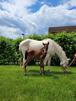
The bond between Pearl and the foal strengthened over the coming days. The foal’s antimicrobial treatment was discontinued, and the supplemental milk offered in a bucket was reduced as the colt was nursing from Pearl. At the time of Hospital discharge, the foal was healthy and active with normal foal behavior.
More than two months following the foal’s birth, he’s reported to be doing well at the barn where he lives with his nurse mare, Pearl.
Comments
Equine dystocia is the ultimate emergency. Experience and expediency are critical for foal survival. From the time that the chorioallantois ruptures, the delivery of the foal should take less than 30 minutes. In fact, prompt intervention is warranted if no progression is made after only 10 minutes of Stage II labor. Delayed delivery increases the likelihood of fetal asphyxia and other neonatal problems associated with hypoxia due to placental separation. Studies have shown that for every 10-minute increase in the duration of Stage II labor beyond 30 minutes, there is a 16% increase in the foal not surviving to hospital discharge.
Options for dystocia resolution involve assisted vaginal delivery, controlled vaginal delivery, cesarean section, and fetotomy. Assisted vaginal delivery is performed with the mare standing. This is what was attempted on the farm by Rouby’s primary veterinarians. This technique is typically successful for simple mutations only (flexed extremities). Controlled vaginal delivery is performed with the mare under general anesthesia and positioned in Trendelenburg position with the hind limbs suspended. This allows for deeper repulsion and can be successful for more difficult mutations (head and neck retroflexion). Because time is so critical for survival of the foal, if the foal is suspected to be alive on hospital admission, assisted vaginal delivery is not attempted for more than 10 minutes before moving on to controlled vaginal delivery.

Likewise, controlled vaginal delivery should only be attempted for 15 minutes before proceeding to a cesarean section if the foal is potentially alive. In Rouby’s case, there was no indication that the foal was alive, and statistically speaking, with the amount of time that had passed since the initiation of Stage II labor, the probability of a viable foal was virtually zero. Because of this, controlled vaginal delivery was attempted for a much longer period. Additionally, the cost of a cesarean section on a horse and the postoperative recovery period was also considered as Rouby was 18 years old and was not going to be bred again.
Because Rouby had been in labor for more than five hours by the time that the foal was delivered, the team was utterly shocked that the foal was alive and even more impressed by how well he did following delivery. Despite having all the odds against him, this colt was one of the healthiest foals the team has seen this year. Surprises like these are always welcome!

