What the new scanner does
- Uses newly developed X line high-performance objectives to deliver optimal digital image quality for quantification through better resolution, flatness, and a wider field of view
- Provides greater flexibility for multiple applications on individual tissue sections; these applications include brightfield, phase contrast, darkfield, polarization, and fluorescence
- Improved slide loader design and capacity holds up to 35 sample trays with a maximum capacity of 210 glass slides; this doubles the old scanner’s capacity
- Can scan 100 slides in a single overnight session
- Allows for whole slide scanning
- Can digitize images of entire glass slides for better storage and sharing; this means no more boxes of slides, just hard drives
- Offers a variety of other capabilities like AI-powered pathology recognition, quantitative pathology, and different types of microscopy using OLyVIA software
- Magnifies up to 40x
- Allows clinical pathologists to read blood slides using oil to further increase magnification to 60x
In 2010, an Olympus VS110 Virtual Microscopy System was installed at MSU’s College of Veterinary Medicine. That system provided state-of-the-art microscopic imaging and digitization of whole glass slides that contained tissue sections from laboratory or domestic animals. At the time, MSU was one of only a few academic institutions in Michigan or elsewhere with this imaging technology. More than a decade later, the digital slide scanner now requires more maintenance--service calls and replacement parts--due to an ever-growing workload. This reduced availability and increased demand led to delays in pathology reports and scientific publications critical for research.
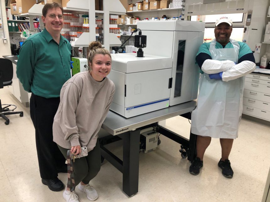
Now, a brand new, updated Olympus SLIDEVIEW VS200 Research Slide Scanner has been installed in the Laboratory for Toxicologic and Environmental Pathology in MSU’s Food Safety and Toxicology Building. But this new scanner won’t only help balance workload—it offers much more.

“The new VS200 scanner greatly improves our microscopic imaging capabilities for a variety of histologic tissue samples garnered for biomedical research,” says Dr. Jack Harkema, director of the Laboratory and University Distinguished Professor. He notes that the scanner’s improved image quality, microscopic imaging capabilities, and data storage and sharing will streamline work.
The new scanner also allows for a variety of image adjustments and measurements. With improved optics, the images have greater resolution, which is necessary for identifying signs of disease, assessing lesion severity, and making a diagnosis. “When thinly cut tissue sections are laid on glass slides, they aren’t perfectly flat. The digital slide scanner automatically adjusts as it scans so everything is in perfect focus within a single plane,” says Harkema.
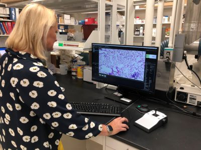
The new scanner’s software boosts capabilities even more by connecting the researcher’s computer to their microscope. Users can capture and download high-quality images for scientific reports and publications.
Once finished “reading” digitized images, users can save them onto a hard drive or other storage device for transport and sharing. Harkema notes that digital pathology has been especially helpful during the pandemic. “If I’m working on a research study or assisting on a specific case, the VS200 Slide Scanner allows me to share scans of whole glass slides with others, almost anywhere, as quickly as I can upload the files, send them electronically, or set up a Zoom call,” says Harkema.

To obtain the scanner, a multidisciplinary group of researchers at MSU submitted a detailed proposal to Dr. Douglas Gage, vice president of Research for the University. The College of Veterinary Medicine made a large financial commitment for the purchase of the new research slide scanner, but several other biomedical researchers and research units at MSU contributed as well, including the Office of Research and Innovation.
“This new scanner will benefit so many researchers at MSU. It’s a powerful tool to have at our disposal, and as producers of critical biomedical and environmental research, we’re already putting it to good use,” says Dr. Dalen Agnew, professor and chair for MSU’s Department of Pathobiology and Diagnostic Investigation at the College of Veterinary Medicine.
A selection of slide scanner users
- Dr. Jack Harkema, University Distinguished Professor, College of Veterinary Medicine
- Dr. James Pestka, University Distinguished Professor, College of Agriculture and Natural Resources
- Dr. James Wagner, Associate Professor, College of Veterinary Medicine
- Dr. Dalen Agnew, Professor and Chair, College of Veterinary Medicine
- Dr. Tim Zacharewski, Professor, College of Agriculture and Natural Resources
- Dr. James Luyendyk, Professor, College of Veterinary Medicine
- Dr. Linda Mansfield, University Distinguished Professor, College of Veterinary Medicine
- Dr. Cheryl Rockwell, Associate Professor, College of Osteopathic Medicine
- Dr. Greg Fink, Professor, College of Human Medicine
- Victoria Watson, Assistant Professor, College of Veterinary Medicine
- Cheryl Swenson, Associate Professor, College of Veterinary Medicine
