History and Presentation
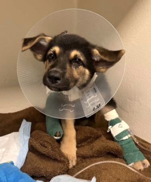
Max, an 8-week-old German Shepherd, presented to the Small Animal Emergency and Critical Care Service at the MSU Veterinary Medical Center after being accidentally stepped on while playing with an 80-pound dog at home. He was non-ambulatory on his right pelvic limb, with signs of pain that included a refusal to eat or drink.
Diagnosis
A radiograph confirmed that Max had suffered a closed, complete, minimally-displaced mid-diaphyseal spiral fracture of his right tibia, with an intact fibula. Due to the type of fracture Max sustained, surgery was recommended to stabilize the tibia to prevent further displacement while it healed, and allow him a quicker return to function than bandaging would have provided.
Treatment and Outcome
Since the fracture was minimally displaced, Dr. Marturello performed his repair minimally invasively through two small incisions. An epiperiosteal tunnel was created using a 14-hole 1.5/2.0mm cuttable plate, which was passed proximal to distal through the two incisions and positioned on the medial aspect of the tibia. The plate was affixed with one proximal and one distal non-locking 2.0 mm screws. C-arm fluoroscopy was used to confirm appropriate implant placement and avoidance of the growth plates. The incisions were routinely closed in layers with 3-0 PDS and 4-0 Monocryl.
Post-operative radiographs showed anatomical alignment of the limb with excellent implant positioning. Post-operatively, the owner was given the option of a light supportive bandage for 3-4 days, or to take Max home without one. Due to his rambunctious nature and the 1:1 screw configuration, she elected for the added support over the weekend. Max returned 3 days later for bandage removal.

Comments
The prognosis for Max to return quickly to normal function was excellent, particularly with appropriate post-operative management and exercise restriction. Since the repair was performed minimally invasively, and the chosen fixation was compliant in accordance with the technique (elastic plate osteosynthesis), Max was anticipated to heal within a week as long he was adequately activity restricted. Maintaining ideal body weight is also imperative in the management of orthopedic injuries.
As the joint was not involved in Max’s fracture, osteoarthritis secondary to this surgery was considered unlikely. Potential complications could have involved infection, delayed union of the fracture site, implant failure, incision dehiscence, or seroma formation.
Fortunately, Max’s owner reports that he has recovered well. In young dogs, a robust and quicker healing process is generally expected as compared to an adult dog.
Below: The below video shows Max walking and bouncing at his one-week-post-op recheck appointment. Radiographs performed at this visit showed that Max’s fracture had healed without complications, and Max was allowed to return to normal activity levels one week after surgery.


